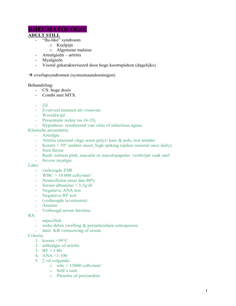
RHEUMATOLOGIE
ADULT STILL
- “flu-like” syndroom
o Keelpijn
o Algemene malaise
- Artralgieën – artritis
- Myalgieën
- Vooral gekarakteriseerd door hoge koortspieken (dagelijks)
overlapsyndromen (systeemaandoeningen)
Behandeling:
- CS: hoge dosis
- Combi met MTX
- ZZ
- Evenveel mannen als vrouwen
- Wereldwijd
- Presentatie ziekte tss 16-35j
- Hypothese: resulterend van virus of infectieus agens
Klinische presentatie:
- Artralgie
- Artritis (meestal oligo soms poly): knie & pols, rest minder
- Koorts > 39° sudden onset, high spiking (spikes meestal once daily)
- Sore throat
- Rash: salmon pink, macular or maculopapular, verdwijnt vaak snel
- Severe myalgie
Labo:
- verhoogde ESR
- WBC > 10 000 cells/mm³
- Neutrofielen meer dan 80%
- Serum albumine < 3,5g/dl
- Negatieve ANA test
- Negatieve RF test
- (verhoogde levertesten)
- Anemie
- Verhoogd serum ferritine
RX:
- aspecifiek
- weke delen zwelling & periarticulaire osteoporose
- later: KB vernauwing of erosie
Criteria:
1. koorts >39°C
2. arthralgie of artritis
3. RF < 1:80
4. ANA <1:100
5. 2 vd volgende:
o wbc > 15000 cells/mm³
o Still’s rash
o Pleuritis of pericarditis
1
o Hepatomegalie of splenomegalie of gegeneraliseerd
o lymphadenopathie
ARTERITIS TEMPORALIS = giant cell arteritis
- geassocieerd met PMR
- hoofdpijn, soms tot in haren
- giant cell arteritis – biopsie
Behandeling:
1jaar medrol 32mg (CS)
o Neveneffecten
o Oedemen
o Huidbroosheid
o Haaruitval
overlapsyndromen
diagnose & behandeling niet op tijd blind worden pt
- incidentie stijgt met leeftijd (bijna enkel > 50j)
- vrouw > man
- hoogste prevalentie in Scandina & N Eur
- mononucleair cel infiltraat door alle lagen van mid-sized arterie struct verandering
- non thrombotic luminal occlusion
- genetisch: HLA-DR4
clinical features:
1. tekens van vasculaire insufficiëntie
2. tekens van systemische inflammatie
cranial arteritis
- hoofdpijn: throbbing, Sharp or dull
- schedel tenderness
- ischemische optische neuropathie vision loss: sudden, painless and usually perman
- kaak claudicatio (m masseter en temporalis) + mog trismus
- mog claudicatio van tong en pijnlijke dysfagie
- CNS ischemie
- PMR
- Directe respons op CS
- Pulses zijn gereduceerd of afw
- Resp symptomen
- Carotis/vertebro aa TIA of CVA
Labo:
- sterke verhoogde acute fase prot: CRP, IL-6
- sterk verhoogde ESR
- verhoogde bp
- mog abnl leverfct testen: alkaline fosfatase
criteria:
1. age bij ziekte onset: >50j
2. nieuwe hoofdpijn
3. temporal arterie abnormaliteit
4. verhoogde ESR
5. abnormale arterie biopsie
2
DERMATOMYOSITIS
- proximale spierzwakte
belangrijkste klacht
- last met boodschappen dragen
- artralgieën, artrtis: milde oligoartritis
gewrichtspijn – geen spierpijn!
- associatie met neoplasie (secundair aan tumor)
- huidaantasting:
o spier & huid aangetast
o huidrash
o ‘getron? Nodules’
o Oedeem rond ogen
- alles kan mee aangetast zijn
inflammatoire spierziekten
Bloedbeeld:
- gestegen bloedenzymen (CK)
-
vrouw/man = 2/1
raciale verschillen
autoimmuunverschijnsel
associatie met andere autoimmuunziekten
myositis-specifieke autoantibodies = MSA
o antisynthetase (anti-Jo-1)
o anti-Mi2
- myositis-geassocieerde autoantibodies
- HLA-DR3 – HLA-138 – DR3 – DR6
Clinical features:
- symmetrische proximale spierzwakte
- systemische symptomen
o moeheid ( abnl spier E metabolisme & veranderde excercise capaciteit)
o ochtendstijfheid
o anorexia
- EMG
- Spierbiopsie: inflamm veranderingen
o Perifascicular atrofie
- Rash (oa pathogn: Gottron’s papuli)
- Periungual teleangiectasias
- Nail-fold capillary changes
- Raynaud’s fenomen
- Juvenile dermatomyositis
o Vasculitis
o Ectopic calcification
o Lipodystrofie
o Spierzwakte
o GI ulcerations
labo:
- verhoogde serumenzymen afgeleid van skeletspieren
3
o creatinine kinase
Criteria: polymyositis en dermatomyositis:
1. symmetrische weakness
2. spierbiopsie bewijs
3. verhoging van spierenzymen (in serum)
4. EMG bewijs
5. dermatologische kenmerken
Bevindingen die dermatomyositis of polymyositis uitsluiten:
- bewijs voor centrale of perifere neurologische disorder
- spierzwakte met een traag progressief unremitting verloop en een pos fam gesch of
gastrocn vergroting spierdystrofie
- biopsie bewijs voor granulomateuse myositis (sarcoïdose)
- infecties
- recent gebruik van medicijnen en toxines (clofibrate en alc)
- rhabdomyolyse
- metabole stoornissen
- endocrinopathiën
- endocrinopathiën
- myastenia gravis
ENTEROPATHISCHE ARTRITIDEN
- vooral OL, knie en enkel
- vrij acuut
- migratoir
- asymmetrisch
- oligoarticulair, perifeer en/of axiaal
- inflammatoir gewrichtsvocht (tot 50 000 cells/ml, PMNs)
- extra-intestinale manifectaties
o pyoderma gangrenosum
o afteuze stomatitis: mondaftose
o uveitis
o erythema nodosum
rode zwellingen OL, rood, verheven, warm, pijn
- artritis kan voorafgaan aan de GI symptomen
Etiologie:
- Crohn, colitis ulcerosa
- Infectieuze:
o Salmonella
o Shigella
o Campylobacter
- Microscopische/idiopathische colitis
- Whipple
- Intestinal bypass
FIBROMYALGIE
Cervicale pijn tender points
-
diffuse pijn voor verschillende jaren
4
-
subjectieve klachten:
o moeheid
o geheugenproblemen
o slaapstoornissen
o irritable bowel symptoms
- familiale aggregatie
- triggers stressors:
o fysiek trauma
o infecties
o emotionele distress
o endocriene aandoening
o immuun stimulatie
Criteria:
1. history of wide spread pain
Definitie:
Pain is considered widespread when all of the following are present:
- pain in the left side of the body
- pain in the right side of the body
- pain above the waist
- pain below the waist.
In addition, axial skeletal pain (cervical spine or anterior chest of thoracic spine or low back)
must be present. In this definition, shoulder and buttock pain is considered as pain for each
involved side. “Low back” pain is considered lower segment pain.
2. pain in 11 of 18 tender point sites on digital palpation
Definitie:
Pain, on digital palpation, must be present in at least 11 of the 18 tender point sites
JICHT
- diffuse pijn
- diffuse zwelling
- urinezuur
- DIP mee aangetast
- Monosynovitis
- Artritis mutilans
- Radiologie:
o Weke delen zwelling
o Tofi
o Bony erosions met sclerotische randen en overhangende randen
o Gewrichtsspleet is bewaard
- Tophi
- Acuut ontstane pijn
- Gewrichtspuntie: kristallen in + strongly inflammatory (tss 20 000 en 50 000wbc)
- Begin: acute aanval inflamm monoarthritis MTP (knie ook vaak)
- Volgende aanvallen:
o gebeuren frequenter
o oligo tot polyarticulair
o duren langer
kristalarthropathie
Etiologie: metabool
5
Bloedbeeld:
- RF: 5à10% pos
- hyperuricemie
Behandeling:
verlagen urinespiegel
o allopurinol
o probenecid
-
-
-
-
-
-
supersaturatie van urinezuur in ECF
o overproductie > 800mg urinezuur geëxcreteerd
o onderexcretie (90%)
o combinatie (alcohol)
volw mannen: piek in 5de decade
(samenhang met hypertensie?)
Associatie met chronische nier insufficiëntie
Genetische factoren
Omgevingsfactoren:
o LG
o Dieet
o Levenstijl
o Sociale klasse
o Hemoglobine concentratie
3stadia:
1. asymptomatische hyperuricemie
2. acute intermittente jicht
3. chronische tophaceouse jicht
jicht aanval: begint zacht mr wordt enorm pijnlijk
o warmte
o zwelling
o erytheem (lijkt op cellulitis)
o pijn
8 à 12u durend
Verschil tss ‘petit attacks’ en ‘severe attacks’
meestal MTP1 (ook midfoot, enkel, hiel, knie; beperkt: pols, vinger en
elleboog)
Systemische symptomen:
koorts
chills
malaise
mog subklinische inflammatie
chronische tophaceous jicht
o 10jaar of meer na acute intermittente jicht
o Periodes tss aanvallen nt meer pijnvrij
o Tofi: vinger, pols, oor, knie, olecranon bursa, drukpunten
mog vorming Heberden’s noduli
o Gewrichten persistent oncomfortabel & gezwollen (verergert bij aanval)
o Diffuse en symm aantasting kleine gewrichten handen en voeten
Associaties:
6
o
o
o
o
RX:
-
Nierpathologie
Hypertensie
Obesitas
hyperlipidemie
early: unremarkable
acute jicht: weke delen zwelling rond aangedane gewricht
later: bot en gewricht misvorming kristaldepositie
assymm
in feet, hands, polsen, elbows, knees
Erosies licht verwijderd van het gewricht: atrofisch en hypertrofisch
overhanging edge
labo:
- hyperuricemie: 2SD boven gem (boven 7,0 mg/dl voor mannen, 6,0 mg/dl voor
vrouwen) max oplossend vermogen serum is 6,7mg/dl
- leucocyten: gem 15 à 20 000 cells/mm³
Clinical manifestations:
- recurrent attacks of articular and perarticular inflammation
- accumulatie van articulaire, osseuze, weke delen en KB Kristal deposits
- urinezuurcalcinuli in de urinaire tractus
- interstitial nefropathie met renale fct verstoring
Diagnose:
- acute monoartritis
- verhoogde urecemie
- sterke verbetering door colchicine
MIXED CONNECTIVE TISSUE DISEASE
- systeemsclerose – systeemsclerose – myositis
- spierkrachtvermindering
overlapsyndromen (systeemaandoeningen)
Bloedbeeld:
- ANF: typisch anti-RNP
- CK gestegen
-
één vd pediatrische reumatoïde ziekten
variant van SLE
Raynaud
Hypergammaglobinemie
ANA +
RF +
Antibodies voor Sm –
Antibodie voor U1 ribonucleoprot +
Nail-fold capillary changes
Gootren’s papules
OSTEOARTROSE = ‘osteoartritis’
- Middle-aged en oude mensen
- beenderige/hard zwelling
7
-
-
-
-
typisch PIP’s en DIP’s
knie met of z meniscus of ligamentair probleem
oudere patiënt
mechanische pijn, belastingsgebonden
o vermindert met rust
o gn nachtelijke pijn
o geen nachtelijke stijfheid
o wel startpijn
o pijn afkomstig van subchondrale bot en andere intra- en periarticulaire
structuren zoals synovium, menisci en ligamenten
acute flares met een inflammatoire component en zwelling kunnen voorkomen
weinig of geen synovitis
pijn komt progressief op over weken
acute monoartritis met chondrocalcinose
niet-inflammatoir
botscan: weinig nut
chronische oligo/polyartritis
polyartralgie (&milde synovitis)
non-inflammatoir
beperkte functie aangetast gewricht
KO
o Pijn bij actieve en passieve beweging
o Lokale tenderness
o Zware gevallen: crepitus
o Gewrichtsvergroting:
Hydrops
Synovitis
Osteofytosis
Bone remodelling
o Spieratrofie (secundair dr disuse)
o Secundaire asdeviaties
o Retropatellar OA: pijn rond patella – erg bij trappen
Punctie:
o weinig abnl <2000 wbc
o calcium pyrofosfaat of apatite kristallen: regelmatig gezien
flexie: asymmetrisch
discus degenereert: spleet verkleint draagopp vergroten door horizontale osteofyten
C5-C7 nekpijn
Patroon van gewrichtsaantasting:
o Wel:
DIPs, PIPs, CMC I (duimbasis)
MTP I
Knie-heup
Acromioclaviculair
Apofysaire gewrichten vd cervicale en lumbale WZ
o Niet:
MCPs (indien wel: hemochromatose)
Polsen
Ellebogen
Glenohumeraal
8
-
-
-
Enkels
MTPs II-V
Radiologie:
o Gewrichtsspleetvernauwing (compartimenteel)
o Subchondrale sclerose
o Osteofyten
o Geoden
o Vacuum sign (discopathie)
CT en MRI: vroeger afwijkingen opsporen
Trofostatisch syndroom?:
o Anterolisthesis/posterolisthesis
o Apofysaire gewrichtsaantasting
Radiculopathie
Jonge persoon: associatie met meniscus of chondraal/osteochondraal probleem of
defect MRI, CT-arthrografie en arthroscopie
etiologie: degeneratief
- erfelijk
- biomechanische factoren
o traumata
- celbiologische factoren
- leeftijd
- overgewicht
bloedbeeld:
- RF: 5à10% pos
- Normaal
- Soms lichte stijging CRP
- Licht stijging ESR
Behandeling:
non-medical
- educatie
- gewichtsverlies
- goede spiersterkte
medical
- paracetamol
- NSAID
- Topical anti-inflammatory treatment
- Voedingssupplementen:
o Glucosamine
o Chondroïtine sulfaat
- (intra-articulair: CS en hyaluronzuur)
chirurgie
definitie :
OA is het resultaat van zowel mechanische als biologische gebeurtenissen die het normale
evenwicht verstoren tussen degradatie en synthese van articulair kraakbeen en subchondraal
bot.
als monoartritis :
9
- suddenly worsening pain and swelling in a single joint
- nieuwe pijn als gevolg van overbelasting of mineur trauma
- ouderen: spontane osteonecrose (vnl in knie)
- door penetrerende wonden van doornen, hout of ander vreemd materiaal
algemeen:
- vaak cervicale en lumbale WZ
- meest: gewichtsdragende gewrichten in been en bep gewrichten in hand
- lokale factoren:
o overgewicht
o injury and occupation
o developmental deformities
genu varum
genu valgum
congenitale heupluxaties
slipped capital femoral epifysis
Legg-Calvé-Perthes
Acetabulaire dysplasie
o Kracht quadriceps
o Knielaxiteit
o Schade aan lokaal kraakbeen
- Systemische factoren:
o Bescherming van oestrogeen
o Genetische voorbeschiktheid
o Meer bij Afr-Am vrouwen
o Lage inname van vit C en D
o Metabole en endocriene ziekten
- Clinical features:
o Gewrichtspijn:
gradueel of insidieus qua begin
mild tot moderatie
betert met rust w later persistent
erger met regenweer
o Tenderness
o Beperking van beweging
o Crepitus
o Occasioneel effusion
o Variabele graad van inflammatie
o Stijfheid & beperkte fct gewricht
- RX:
o Bony proliferation aan gewrichtsmargin
o Asymm gewrichtsspleet
o Subchondrale bot sclerose
o Verkalking kraakbeen
- MRI:
o KB direct in beeld
o Osteofyten
o KB verlies
o Subchondrale cysten
o Meniscal en ligamenteuze abnormaliteiten
- Labo:
10
o
o
o
o
o
Routine tests: nl
Ca – Fosfaat - Mg – TSH
Voor NSAID te geven: Hb – creatinine – K- aspartaat aminotransferase
ESR nl voor leeftijd
WBC < 2000 cells/mm³
OSTEOPOROSE
- mechanische pijn
o diffuus
o unilat
o variabele onset
o ochtendstijfheid <30min
o activiteit verergert symptomen
o rust verbetert symptomen
o milde nachtelijke pijn
- weinig of geen synovitis
- geen systeemaandoeningen
- rugpijn: fracturen
- osteoporose doet geen pijn!
Alleen fracturen/indeukingsfracturen
- BMC meting
o T-score vs jong volwassene: < -1 = osteopenie
o Z-score vs age-matched: <-1 = osteoporose
o Waar?
Lumbaal (trabeculair)
Femoral (corticaal – trabeculair)
Voorarm (cortical)
-
-
<-2,5 = osteoporose
types osteoporose
o type I
55-75j
6vrouwen/1 man
Trabeculair > cortical botverlies
Anatomische plaats: radius = WZ
Etiologie: tekort oestrogenen
o type II
70-85
2vrouwen/1man
Trabeculair = cortical botverlies
Anatomische plaats: femur
Etiologie: veroudering
Risicofactoren
o Familial
o Tengere structuur/bouw
o Vroegtijdige menopause
o Sedentair leven
o Lage kalk inname
o Sigaretten/tabak
o Alcohol (>2drinks/dag)
o Koffie (>2 tassen/dag)
11
o Medicatie: CS, I-thyroxine
Etiologie:
metabool
Behandeling: valpreventie
-
-
lage botmassa en microarchitecturale deterioratie
skeletale fragiliteit
oudere populatie; vnl white vrouwen
leidt tot breuken
klassieke OP breuken:
o heup
o vertebra
o pols
osteopenie: BMD tss 1 en 2,5 SD onder gem & OP: BMD > 2,5 SD onder gem
o DXA meting: dual X-ray absorptiemeter
acute pijn bij osteopor fractuur:
o intense, gelokaliseerde pijn
o gereduceerde WZ beweging
o blijft voor 4 tot 6weken
verlies in hoogte: progressieve kyfose / flattening lumb lordose
rugpijn
meerdere indeukingen: verterings- en AH problemen
PMR = polymyalgia rheumatica
- chron oligo/polyartritis
- polyartralgie (& milde synovitis)
- oudere patiënt
- uitgesproken inflammatoire gordelpijnen
o schouderpijnen, moeilijk rechtkomen
o bekkengordel
- transiënte milde synovitiden (knie, pols)
- spectaculair antwoord op CS
- associatie met arteritis temporalis
- arteritis
- vasculitis
- cervicale pijn: stijfheid, pijn
= systeemaandoening
overlapsyndromen
bloedbeeld:
- hoge sedimentatie (>40 mm)
- gn stijging CRP of ERP
uitsluitingsdiagnose!!!
-
één vd vasculatiden
pijn en stijfheid voor de laatste 4weken: = gordelpijnen
o nek
12
o schoudergordel
o pelvische gordel
- tekens van systemische inflammatie:
o malaise
o gewichtsverlies
o zweten
o laaggradige koorts
- upregulatie acute fase proteïnen
- zeer gevoelig aan CS therapie
- als bloedvatwand inflammatie aanw GCA
- meer vrouwen dan mannen
- zeer onwsl bij pt < 50j
- hoog risiko populatie: Scand & Noord Europa
- sudden onset van intense inflammatie
- genetisch: HLA-DR4
clinical features:
- aching and pain in spieren van:
o nek
o schouders
o lage rug
o heupen
o dijen
o (trunk)
- Pyrexia en chills
- Diffuse oedeem van handen en voeten
- Reageert prompt op therapie
- ZOEK NAAR VASCULAIRE PROBLEMEN!!!
labo:
- verhoogde ESR
- verhoogde CRP
- anemie
POLYMYOSITIS
- proximale spierzwakte
belangrijkste klacht
- last met boodschappen dragen
- artralgieën, artrtis: milde oligoartritis
gewrichtspijn – geen spierpijn!
- associatie met neoplasie (secundair aan tumor)
inflammatoire spierziekten
Bloedbeeld:
- gestegen bloedenzymen (CK)
-
vrouw/man = 2/1
raciale verschillen
autoimmuunverschijnsel
associatie met andere autoimmuunziekten
13
-
myositis-specifieke autoantibodies = MSA
o antisynthetase (anti-Jo-1)
o anti-SRP
- myositis-geassocieerde autoantibodies
- HLA-DR3 – HLA-138 – DR3 – DR6
Clinical features:
- symmetrische proximale spierzwakte
- systemische symptomen
o moeheid ( abnl spier E metabolisme & veranderde excercise capaciteit)
o ochtendstijfheid
o anorexia
- EMG
- Spierbiopsie: inflamm veranderingen
o Verschillende stadia van necrose en degeneratie
o Inflamm celinfiltraten
o Vernietigde vezels w vervangen dr vet en fibrotisch weefsel
- Onset over 3 à 6maand
- Meest:
o Schoudergordel
o Pelvic gordel
o nekspieren
- dysphagia door oesofagale dysfunctie of cricopharyngeal dysfunctie
- myalgie an arthralgie
- soms Raynaud
- mog periorbital oedeem
- pulmonaire en cardiale manifestaties:
o “velcro-like” crackles
o Aspiratie pneumonie
o Asymptomatische ECG abnormaliteiten
o Supraventiculaire aritmie
o Cardiomyopathie
o Congestive hartfalen
- EMG:
1. verhoogde insertionele activiteit, fibrillaties, scherpe pos waves
2. spontane, bizarre, high-frequency discharges
3. polyphasic motor-eenheid potentials of low amplitude and short
duration
labo:
- verhoogde serumenzymen afgeleid van skeletspieren
o creatinine kinase (nt over ganse verloop ziekte)
o aldolase
o aspartate aminotransferase AST
o alanine aminotransferase ALT
o lactaat dehydrogenase LDH
- verhoogde ESR
Criteria: polymyositis en dermatomyositis:
1. symmetrische weakness
2. spierbiopsie bewijs
3. verhoging van spierenzymen (in serum)
14
4. EMG bewijs
5. dermatologische kenmerken
Bevindingen die dermatomyositis of polymyositis uitsluiten:
- bewijs voor centrale of perifere neurologische disorder
- spierzwakte met een traag progressief unremitting verloop en een pos fam gesch of
gastrocn vergroting spierdystrofie
- biopsie bewijs voor granulomateuse myositis (sarcoïdose)
- infecties
- recent gebruik van medicijnen en toxines (clofibrate en alc)
- rhabdomyolyse
- metabole stoornissen
- endocrinopathiën
- endocrinopathiën
- myastenia gravis
PSORIASIS ARTRITIS
- intermittent acuut oligo/polyartritis
- polyartritis – polysynovitis
- chronisch
- vaak ook DIP’s
- knie mogelijk eerste symptoom
- gekarakteriseerd door botaanmaak en osteolyse thv pees, lig en kapselinserties
- joint involvement:
o asymm oligoarticular disease
>50%
DIPs and PIPs of hands and feet, MCPs, MTPs, knees, hips and ankles
Predominantly the joints of the lower limbs
o predominant DIP involvement
5à10%
DIPs
o arthritis mutilans
= zéér snelle destructieve evolutie
5%
DIPs, PIPs
o polyarthritis “rheumatoid-like”
15à25%
MCPs, PIPs and wrists
o axial involvement
20à40%
Sacroiliac, vertebral
- Psoriasis:
= actuele of vroegere psoriasis gediagnosticeerd door een arts
o Vooraf: 67%
o Parallel of na: 33%
- Olievlekken op nagel
- Vnl huidletsels thv drukplaatsen
- Articulaire aantasting:
o Sausage digit
o Enthesitis
o Osteoperiostitis
15
-
-
o Oligoartritis grote gewrichten
o Polyarticulaire artritis
o Artritis mutilans
o Sacroiliitis
o Spondylitis
o Artritis DIP
Extra-articulair:
o Uveïtis
o Subklinische darminflammatie
o (coeliakie)
Prognose: minstens 20% ernstige destructieve artritis met irreversibele misvormingen
Radiologische laesies: pencil in cup lesion subluxatie
Verschil met RA:
o Asymmetrische artritis
o Axiale gewrichtsaantasting
o RF afwezig
o Nail pitting
o Sausage digits
o Family history of psoriasis/psoriasis artritis
o Radiologisch: belangrijke remodelling, weinig peri-articulaire osteopenie
Etiologie:
multifactorieel
- immunologisch: TNF, PDGF, VEGF, substance P, VIP
- genetisch: HLAsysteem: Cw6, B13, B17, DR7, B27
TNFalfa6c1d3
- vasculair
- omgevingsfactoren: streptococceninf, HIV, enterobacteriën, HepC
- neuronaal: paralyse
bloedbeeld:
- RF: 10à25% pos
- Hoge titers TNFalfa
Behandeling:
- houdings- en lenigheidsoefeningen
- medicatie
o NSAID
o Basistherapie
MTX, salazopyrine
TNF blokkade
Pediatric rheumatic disease: juveniele psoriatic arthritis
- chronische artritis in associatie met psoriasis
- onset < 16j
- rash kan jaren achterblijven
- criteria: definite psoriatic arthritis
o artritis and psoriasis
o of arthritis + 3 vd 4 volgende:
1. dactylitis
16
2. nail pitting of onycholysis
3. psoriasis in fam (1e of 2e gr verwanten)
4. atypische psoriasis rash
o
- Arthritis perifeer
- Geassocieerd met asymptomatische chronische uveïtis
- Flexor tenosynovitis in vingers of tenen (sausage digits)
Algemeen:
- variërende vormen:
o monoarthritis
o asymm oligoartritis
o symm polyartritis
- 0,1% mensen – 5à7% in mensen met psoriasis
- Vnl Caucasians
- Peak of onset: tss 30 en 55j
- Genetische factoren:
o Familial
o HLA-B27 – HLA-B13 – HLA-B16 – HLA-B17 – Cw6 – MICA-002
- Omgevings factoren:
o Streptococci & staphylococci
o Trauma
o HIV
- Immunologische factoren:
o Activatie en expansie van weefsel-specifieke subsets
o Accumulatie van inflammatoire cellen
o Angiogenese
o Relatieve deficiëntie IL-10
- Clinical features:
1. mono- of oligoarthritis met enthesistis resembling reactive arthritis
2. symm polyarthritis reassembling RA
3. predominantly axial disease
spondylitis
sacroiliitis
arthritis van heup of schoudergewrichten
o DIP regelm betrokken
o Arthritis mutilans
o Sacroiliitis
o Spondylitis
o Psoriasis (begin: voor – parallel – na)
o Nagelveranderingen
o Fam gesch met psoriasis of psoriasisarthritis
o Andere manifestaties:
Enthesitis
Ocular involvement: conjunctivitis
Aortic insufficiency
Uveitis
Pulmonary fibrosis
amyloidosis
o dermatologisch:
17
-
sharply demarcated erythematous plaque with a well-marked, silvery
scale
nail involvement:
pitting
onycholysis
transverse depression
cracking
subungual keratosis
…
RX: erosie + bone production
o Fusiforme weke delen zwelling
o Bilat asymm
o Behoud nl minieralisatie
o Dramatisch verlies gewrichtsspleet (vingergw verwijding)
o Pencil-in cup deformatie
o Fluffy periostitis
o Spondylitis
o syndesmofyten
REACTIEVE ARTRITIDEN
= infectie-geïnduceerde inflammatoire synovitis (1-4 weken erna) waarin geen levende microorganisme kunnen aangetoond worden, typisch op een genetische achtergrond (HLA-B27)
peptide-mimicking
- asymmetrische oligoartritis soms erosief bij evolutie naar chroniciteit
- OL, knieën, enkels, voeten
- Enthesitis
- Spondylitis, asymmetrische of unilaterale sacroiliitis
- Synovitis
- Enkele dagen na koorts/GI syndroom ontstaan
- Monoartritis mogelijk
REITER
- polyartritis – polysynovitis
- chronisch
- conjunctivitis
- uretritis
- urogenitaal
- 2 glazenproef
- Keratoderma blenorrhagica: blaren met huidschilferingen
- Radiologie axiaal skelet
seronegatieve spondylartropathie
Etiologie:
Reiter syndroom
o Urethritis
o Artritis
o Conjunctivitis
Urogenitaal
o Chlamydia (PCR urine)
18
Enterogeen
o Shigella
o Salmonella
o Yersinia
o campylobacter
andere
o Borrelia (Lyme): vroegtijdige tekens
Klinische presentatie
- onset: 13-60j: gem 26j
- 87% zijn mannen
- 8/10 Whites – 2/10 Afr Am
- 2 of 4 weken na infectie (GI of veneraal)
- Gewrichten: gezwollen – warm – tender – pijnlijk bij act/pass beweging
+ eytheem
- Sausage digits
Clinical features:
- nongonococcal urethritis
- conjunctivitis
Peripheral arthritis
- nooit enkel BL
- enkel OL 39%
- beide 61%
- meest frequent: knie en kleine gewrichten voet
Affected joints:
- gem 4,5
- sausage digits
- hielpijn (mog met calcificatie)
Spinal arthritis:
- low back pain
- (sacro iliitis - spondylitis)
Mucocutaneouse lesies
- keratoderma blenorrhagicum
- circinate balanitis
- oral ulcers
- nail changes
Andere kenmerken:
- koorts
- gewichtsverlies
- uveitis
Labo:
- anemie
- leucocytose verhoogd
- thrombocytose
- verhoogde ESR
- RF –
- HLA-B27 +
- Acute fase proteïnen zijn abnl
- Serum globulines (vnl IgA) gestegen
- ANA –
19
Synovial fluid:
sterk inflammatoire veranderingen: turbid – slechte visceusiteit – slechte mucine clot test
mr glc niv is niet gedaal ( true septic arthritis)
mog Reiter cells
Oorz:
- diarrheal illnesses: Shigella – Salmonella – Campylobacter
- veneral infectie: Chlamydia trachomatis
JCA
- acute mono/oligo/polyartritis als presenteervorm
- pauciarticular: enthesitis of the tuberositas tibiae in 10%
bloedbeeld:
- RF 10à25% pos
SPONDYLOARTROPATHIE
- extra-articulair
o mondaften
o conjunctivitis
o uveitis
o psoriaris
o uretritis
o diarree
o erythema nodosum
- acute oligo/polyartrtis (Reiter, psoriasis)
etiologie: immuunaandoening
criteria:
inflammatoire spinale pijn
Of
synovitis; asymmetrisch of predominant in OL
Met 1 of meer vd volgende:
- positieve familiegeschiedenis
- psoriasis
- IBD
- Urethritis, cervicitis of acute diarree binnen de maand voor arthritis
- Bilpijn verspringend tss rechtse en linkse gluteale areas
- Enthesopathie
- Sacroiliitis
Pediatric:
1. juvenile ankyloserende spondylitis
2. reactieve artritis
3. psoriatic arthritis
4. arthropathie met IBD
a.seronegatieve enthesopathie
b.seronegatieve arthopathie
- HLA-B27 +
- familie gesch van spondyloarthropathie
- definite arthritis (not just arthralgia)
Algemeen:
20
1.
2.
3.
4.
5.
6.
7.
8.
inflammatoire axial spine involvement (sacroiliitis & spondylitis)
asymmetrische perifere arthritis
enthesopathie
inflammatoire oogziekte
overlappende mucocutaneuze kenmerken
RFFamiliale aggregatie
Associatie met bepaalde HLA’s : vnl HLA-B27
ankylosing spondylitis
reactieve arthritis
psoriatic arthritis/spondylitis
arthritis/spondylitis van IBD
juvenile spondyloarthropathie
undifferentiated spondyloarthropathie
acute anterior unveitis
geïsoleerde AV Block
KRISTALARTHRITIDEN
- ouderen: meestal vanaf 30-35j
- pijn komt progressief op over uren
- gewrichtspunctie: groep3 + kristallen in
- CPPD(pseudogout) – hydroxyapatiet – calciumoxalaat – uraat(jicht)
- Predominantly: acute inflamm monoarthritis of recurrente episoden van inflamm
mono/oligoarthritis
Etiologie: metabool
-
CPPD = calcium pyrophosphaat dihydraat crystals (nt enkel in gewrichtsKB) 4%
o Asymptomatic
o Pseudo-gout: presentatie lijkt op jicht
o Lijkend op septische artritis – polyarticular inflamm arthritis - OA
Chondrocalcinose: Ca bevattende kristallen in articulair kraakbeen 50%
PSEUDOGOUT
- acute inflammatoire kristal mono- of oligoarthritis
- vaak in combinatie met chondrocalcinose
- RX: deposities CPPD in het kraakbeen
- Punctie gewrichtsvocht: gepolariseerd licht microscopie
- Knie: meer lateraal compartiment aangetast dan med (OA)
Etiologie:
Vrijstelling kristal in gewricht veroorzaakt aanval
Behandeling:
NSAID
3vormen:
1. hereditary
2. sporadic (idiopathisch)
3. geassocieerd met verschillende metabole ziekten of trauma
21
o hyperparathyroidism
o hemochromatosis
o hypothyroidism
o amyloidosis
o hypomagnesemia
o hypophosphatasia
- geassocieerd met verouderen
- dose-related inflammatory host responses op CPPD kristallen shed van KB weefsels in
synoviale caviteit
- degradatie van KB matrix
- colchicine helpt
- lokale abnormaliteit van overmaat van articulaire anion productie
Synovial fluid:
- verhoogde PP concentratie
- kristalbepaling voor onderscheid met jicht
- culture
Clinical features:
acute pseudogout:
- inflammatie in 1 of meer gewrichten die verschillende dagen tot 2 weken duurt
- onset kan even abrupt en severe zijn als jichtaanval
o spontaan
o geprovokeerd door:
trauma
surgery
severe illness
- in bijna alle gewrichten mogelijk; 50% in knie
- asymptomatisch tss aanvallen
- mog ”pseudorheumatoid” presentatie
o meerdere gewrichten
o symm distributie
o laag-gradige inflammatie
- progressieve aantasting van verschillende gewrichten: knie pols, MCP, heup,
schouder, elleboog, enkel
- symmetrisch
- flexie contracturen
- deformiteit knie (valgus)
- chron: superimposed episodes van pseudo-OA patroon
- neurologische manifestaties mog dr depositie in axiaal skelet (vb acute nekpijn)
- systemische manifestaties tijdens aanval:
o koorts
o leucocytose: 12à15000 cells/mm³
o verhoogde ESR
o verhoging acute fase proteïnen
RX:
- punctate and linear densities in articulair hyalijn of fibrocartilagineuse weefsels
- typische lokalisaties depositie:
o articulair KB
o menisci
o acetabulair labrum
o fibrocartilagineuze symfysis pubis
22
o articulaire discus van pols
o annulus fibrosus in intervertebrale disci
Labo:
- 10% lage titers van RF
Criteria:
I.
Demonstration of CPPD kristallen in weefsels of synoviaal vocht (dr RX distractie
of chemische analyse)
II.
A.identificatie van monoklinische or triklinische kristallen showing weakly
positive or no birefringence by compensated polarized lichrmicroscopie
B.presence van typische radiografische calcificatie
III.
A.acute arthritis, especially van knie of andere grote gewrichten
B.chronische arthritis, especially van knie, heup, pols, carpus, elleboog, schouder
of MCP gewricht, especially als het begeleid is door acute exacerbaties. Volgende
kenmerken helpen bij een differentiatie met OA:
1. uncommon site: pols – MCP – elleboog – schouder
2. RX: radiocarpal of patellofemoral gewrichtsspleetvernauwing
3. subchondrale cyste vorming
4. severity van degeneratie: progressief met subchondrale botcollaps en
fragmentatie met vorming van intra-articulaire radiodense lichamen
5. osteofyt vorming – variabel en inconstant
6. pees calcificaties (triceps, Achilles, obturators)
SYSTEEMZIEKTEN
VASCULITIDEN
Etiologie:
immuunaandoening
-
ingedeeld volgens grootte aangedaan BV
vb PAN voor middle sized vessels
PSS = progressive systemic sclerosis
- chronische oligo/polyartritis
- polyartralgie (&milde synovitis)
SKELETDYSPLASIE
Etiologie:
congenitaal
OI = osteogenesis imperfecto
- lage botdensiteit
Etiologie:
-
congenitaal
osseous, ocular, dental, aural and cardiovascular involvement
type’s:
o typeI: mog klein gestalte met frequente breuken, gewrichtslaxiteit
o typeII: lethal in infancy
o typeIII: severe skeletal deformity, kyphoscoliose, klein, variabele fracturen
o typeIV: cfr I mr z blue sclera en minder breuken
NEOPLASIE
23
-
pijn komt progressief op over weken
diffuse zwelling hand: longtoptumor?
Synoviale biopsie
Etiologie:
primair of secundair
Primaire gewrichtsneoplasie:
pigmented villonodular synovitis
- symptomen: episodic or slowly progressief
- complete bloedtelling & ESR zijn nl
- aspiratie gw: bruin, rood of geel vocht
synovial chondromatosis
solitair intra-articulair lipoma
synovial hematoma
Secundaire gewrichtsneoplasie:
synovial sarcoma
giant cell tumor
CARPAAL TUNNEL SYNDROOM
- neurogene pijn: tintelingen over distributie gebied N medianus
- EMG: verlengde latentietijd
Etiologie: inklemming N medianus
Bloedbeeld: nl
-
paresthesie & numbness in hand
encroaching kanaal compressie N medianus
episodes van brandende pijn of tingling in hand
gevoel handzwelling
soms pijnverspreiding tot boven elleboog
positief teken van Tinel
verlies sensatie
chron: atrofie thenarspieren
oorzaak:
o oedeem
o osteofyten
o ganglia related to tenosynovial sheaths
o lipomata
o anomalous muscles, pezen en BV
ARTRITIS
- inflammatoire pijn
- zwelling
- warmte
- roodheid
- bewegingsbeperking
INFECTIEUZE ARTRITIS
- diffuse zwelling
24
-
-
(sub)acute monoartritis
voorgesch: cystitis, CS spuit, operatie, huidwonden, …
at lower regions or feet
mogelijk koorts
cultuur gewrichtsvocht
punctie gewricht: groep3
biopsie synovium
voorgesch:
o DM
o Onderliggend gewrichtsprobleem
Mog koorts
Bloed, orale en genitale swaps voor cultuur
etiologie:
synovium of synoviaal vocht wordt cultuurmedium voor bacteriën
bacteraemie
uit aangrenzende weefsels
directe inoculatie dr de huid
S aureus
Streptococci
Enterobacteriaceae
H influenzae b (kids)
Neisseria gonorrhoea (volw)
Fungi (imm def, wonden, IV drug gebruik)
behandeling:
- atroscopische lavage
- drainage
- IV AB (educated-best tot kweekresultaat): nt rechtstreeks in gewricht
GONOCOCCEN ARTRITIS
- eerst: polyarticulair en migratoir
- later: monoarticulair
- vaak ook tenosynovitis en huidletsels (erytheem, pustulae)
- acute oligo/polyartritis
-
migratoire arthritis en/of tenosynovitis
met of zonder huid letsels
sexueel actieve volwassene
jong, gezond persoon
vrouw gevoeliger dan man
synovial fluid vaak negatieve & cultuur levert weinig yield: PCR beter
genitourinary tract cultuur pos in 86%
TBC
- acuut chronisch
- infectieuze artritis
- synoviale biopsie
bloedbeeld:
25
-
RF: 10à25% pos
- osteoarticular involvement in 1 à 5% vd mensen met TBC
- tuberculine huid test is pos
- thorax foto’s vaak nl
Typische presentaties:
- spondylitis (Pott’s disease)
o meestal thoracale wervels
o in endemische gebieden: vnl kids en jonge volw
o Westen: volw door reactivatie van slapende foci
o Ant deel wervellichaam -> disc -> spleet vernauwing -> eindplaat destructie ->
collaps = gibbus
o Mog weke delen inflammatie of psoas abcessen
o Mog sinus tracts
o Mog neurologische schade
- monoarticular arthritis van grote gewichts-dragende gewrichten
o vnl heup en knie
o middle-aged of ouder
o vaak onderliggende medische problemen of CS gewrichtsinjecties
o gewrichtspijn en zwelling
o reactivatie van hematogenously seeded focus
o juxta-articulaire OP
o marginal erosies
o spleetvernauwing
o synoviaal vocht: verhoogd aantal wbc – dominantie PMN
- osteomyelitis en dactylitis
o mog zonder gewricht erin betrokken
- bursitis en tenosynovitis
- reactieve arthritis (Poncet’s ziekte)
o during actieve TBC
o handen en voeten
o gewrichtsvocht & weefselstalen zijn steriel
diagnose:
door identificatie in pus – synoviaal vocht – weefsel
LYME
- acuut chronisch
- infectieuze artritis
- prim: transiënte artralgie & huidafwijkingen
- sec: spetische artritis
- acute oligo/polyartritis
- migrerend
- voorgesch:
o erythema migrans
o disseminated lyme in organen
- laat stadium: neurologische & musculoskeletale symptomen
- onbehandeld:
o 50% migratoire polyarthritis
o 10% chonische, intermittente monoarthritis (grote inflamm art effusions)
- Punctie of biopsie synovium:
26
o PCR
etiologie: spirochete: borrelia burgdorferi
bloedbeeld:
- ELISA & western
Behandeling:
AB
Door Borrelia Burgdorferi
1. B burgdorferi sensu stricto
2. B afzelii
3. B garinii
- ovedracht spirocheet door tekenbeet: te vinden in plasma en clinical samples
- erythema migrans
- genetisch: HLA-DR4 en HLA-DR2
- vasculitis perifere neuropathie
Clinical features:
1. early localized
- erythema migrans
- non-specific complaints:
o koorts
o moeheid
o malaise
o hoofdpijn
o myalgie
o arthralgie
- regionale of gegeneraliseerde lymfoadenopathie
- hepatosplenomegalie
- musculoskeletale klachten
2. early disseminated
- cardiac manifestations
o hartblock
o milde myopericarditis
o chronische cardiomyopathie
- neurologic manifestations
o lymphocystic meningitis
o cranial nerve palsies
o radiculoneuritis
3. late
- chronic arthritis
o meestal monoarticulair (knie)
o erosie van KB en bot
- neurologic features: tertiaire neuroborreliose
o encephalopathie
o neurocognitieve dysfunctie
o perifere neuropathie
- lymphocytoma
- acrodermatitis chronica atrophicans
27
-
panniculitis
myositis, tendinitis, bursitis
liver disease
inflammatie van verschillende oogstructuren
splenitis
nt specifieke klachten:
o hoofdpijn
o moeheid
o arthralgie
Labo:
- ELISA nr antibodies voor Borrelia + Western blot
Synoviaal vocht:
- specifieke AB
- wbc 25 000 cells/mm³
- dominantie van PMN
Spinal fluid: dominantie van mononucleairen
FUNGI
- chronische artritis
- synovium biopsie
fungi:
insidious onset
indolent verloop
milde inflammatie
- candidae (albicans)
- coccidioidomycosis
- sporotrichosis
- blastomycosis
- cryptococcosis
- histoplasmosis
cultuur synoviaal vocht of weefsel
aanwezigheid in weefsel
cultuur van gewrichtsvocht of weefsel
synovial smears – cultuur
aantonen organisme in synoviaal vocht of weefsel
cultuur van weefsel of histologisch aantonen van organismen
HEMORRHAGE ARTRITIS
- punctie knie: haemartrose
- door vroeger trauma
- suddenly °
PERIARTRITIS
- electieve drukpijn
TENDINITIS
- electieve drukpijn
ENTHESITIS
= ontsteking aan peesaanhechting
- electieve pijn
in ankyloserende spondylitis
28
SEPTISCHE ARTRITIS
- diffuse pijn
- ontstaan in enkele uren
- puntie: WBC > 50 000 & > 75% PMN
mortaliteit!!!!
risicofactoren:
- host factors:
o diseased of prosthetic gewricht
o ouder dan 80j
o comorbiditeit: DM, levercirrhose, chronisch nierfalen, maligniteiten, AIDS,
heomfilie, transplantpt
o recente gewrichtschirurgie
o huidinfectie
o immunosuppressie
o hypogammaglobulinemia
o hemodialyses
o IV drug gebruik
o Arthrocentesis of gewrichtsinjectie
o wondcontaminatie
Agens:
o S aureus
o E coliPasteurella multocida
o Pseudomonas aeruginosa
o Coagulase-negatieve stafylococcen
o Gram-neg bacillen
o N gonrrhoeae
o mycoplasma
Clinical features:
- meestal monoarthritis (typisch knie)
- koortsig
- toxic appearance
- extra-articulaire infectieuze focus
- arthrocentesis:
o wbc extremely high: enkele duizenden tot 100 000 cells/mm³
o cultuur
o PMN predominantie > 90%
o PCR
Labo:
- bloedkweek
POLYARTRITIS
- tangentiële drukpijn
- vijf of meer gewrichten
NA MAMMECTOMIE
- diffuse zwelling hand
BURSITIS
- knie: net onder patella zwelling: prepatellaire bursitis
29
anserine
iliopsoas
ischial
olecranon
prepatellar
retrocalcaneal
subcutaneous achilles
trochanteric
BAKER CYSTE
= hernia synoviale membraan
Polscyste: assymmetrische gelokaliseerde zwelling
AVASCULAIRE NECROSE
- botscan
- niet-inflammatoir
- chronisch
OSTEOMYELITIS
- botscan
- infectieus proces
- secundaire rugpijn
MENINGOCOC
- acute oligo/polyartritis
PALINDROOM REUMA
- intermittent acute oligo/polyartritis
periodische disorder van ongekende etiologie
Aanvallen:
- ~2dagen
- Monoarticulaire arthritis of periarticulaire weke-delen inflammatie
- Gewoonlijk onregelmatige intervallen
Betrokken gewrichten:
- MCP
- PIP
- Pols
- Schouder
- MTP
- Enkel
Geassocieerde aandoeningen famiale aggregatie met RA
Prognose:
~50% persistent palindromic rheumatism
~33% ontwikkelen RA
POLYARTRITIS NODOSA = PAN
overlapsyndromen (systeemaandoeningen)
- één der vasculitiden: medium-vessel vasculitis
- koorts
30
- gewichtsverles
- abdominale pijn
- polyneuropthie
- nodulaire zwelling op musculaire aa
- infiltratie gevolgd door fibrinoïde necrose
- beperkte associatie met HepB of hairy-cell leukemie
clinical features:
- begin: nt-specifieke symptomen:
o malaise
o moeheid
o koorts
o myalgie
o arthralgie
- predilictie voor bepaalde organen
o huid : livedo reticulairs – subcut noduli – ulcera – dig gangreen
o perifere nn : mononeuritis multiplex wrist of foot drop
sens abnormaliteiten
motor involvement
Multiple axonal neuropathies
o GI: intestinal angina: postpandrial periumbilical pain
o Nier: renal mediated hypertension
o Cardiac lesions: MI of congestief hartfalen
- Diagnose vereist tissue biopsie of angiogram
Labo:
- anemie
- thrombocytose
- verhoging ESR
- microscopische hematuria
def:
necrotiserende inflammatie van medium-sized of small aa die de kleinste BV sparen en nt
geassocieerd is met glomerulonefritis
REUMATOIDE ARTRITIS
- begin: pijn bij mobilisatie
- pijn komt progressief op over dagen/weken
- begin: synoviale zwelling bilateraal pols
- acute oligo/polyartritis
- gradueel ontwikkelend tot chronisch polyartritis
- symmetrisch
- grote en kleine gewrichten
- zelden DIP’s
- wel cervicaal doch niet thoracolumbaal
- piekincidentie: vrouwen in hun 40-50’ger jaren
- verlies KB – laxiteit/disruptie collat & kruisbanden => moeilijk wandelen en trappen
etiologie:
immuunaandoening met systeemweerslag
chroniciteit:
variërende ziekteactiviteit met opstoten en remissies
inflammatoir polysynovitis floride / ‘bulky’
31
synovium = slagveld
inflamm pijnpatroon: nachtelijke pijn, ochtendstijfheid
chronische polyartritis
symmetrisch: grote (>80%) en kleine gewrichten (MTP, PIP, …)
zelden DIP en thoracale WZ
knie: “bulge” sign en verlies van ‘cool patella’ sign
met sterke synovitis: Baker’s cysts
extra-articulair
algemeen
o vermoeidheid
o gewichtsverlies
huid
o nodules: half hard + los liggen, toch meestal gebonden aan BV, thv
drukplaatsen (vb elleboog, pols, hand)
Heberden/Bouchard
Thv Achillespees
o palmair erytheem
o vasculitis
kan verschillende organen aantasten
opzoeken thv nagels: kleine necrotische zone’s, bloedblaartjes
o huidulceraties: moeilijk genezend
o serositis
oculair
o (epi)scleritis
o Conjunctivitis
o uveïtis
o sicca
cardiaal
o pericarditis
o myocarditis
o hartkleppen (nodules)
pulmonair (RX thorax)
o pleuritis
o nodulus
o interstitieel
o interstitial lung disease
neuromusculair
o entrapment
o mononeuritis multiplex (demyelinisatie – ontsteking)
hematologisch
o Felty syndroom (vergroten milt)
o anemie
Andere
o Sjögren: droge mond & ogen
o amyloïdose
Criteria:
1. Morning stiffness in and around joints lasting at least 1hour before maximal
improvement
2. soft tissue swelling of 3 or more joint areas observed by a physician
32
3. swelling of PIP, MCP or wrist joints
4. symmetric arthritis
5. subcutaneous nodules (laattijdig teken)
6. positive test for RF
7. radiographic erosions or periarticular osteopenia in hand or wrist joints (zeer laat)
4 vd 7 positief RA
Handdeformaties:
- zwanehalsmisvorming
- Boutonnière deformatie
- Polscyste
RX:
vroege periarticulaire osteoporose
gewrichtserosies en gewrichtsspleet vernauwing
dan dislokaties
Echo:
joint effusion
synovial hypertrophy
vascularity
MRI:
aspecifiek botoedeem
Cervicale wervelzuil: C1-C2
Afstand dens-atlas > 3mm
Afschuivingen wervels myelomalacie
GEEN HYPEREXTENSIE/COMPRESSIE MANEUVER THV CERVICALE WZ
Inflammatoir gewrichtsaandoening lage botdensiteit
Bloedbeeld: inflammatoir
- sedimentatie
- CRP
- Anemie (inflammatoire vorm)
- Trombocytose
- Laag serum ijzer
- RF:
o RA latex
o Waaler Rose (meer specifiek)
- Anti-CCP antibodies (cyclisch gecitrillineerde peptiden)
- Mogelijk ANF
Behandeling RA:
Nt-medicamenteus:
o Educatie
o Physical therapy
o Surgical interventions
o Occupational therapy
pijn & inflammatie
o selectieve COX-2 antagonisten?
o CS
33
-
-
o Cytokine blokkade
vrijwaren kracht en mobiliteit
vrijwaren quality of life
beïnvloeden ziekte evolutie en duur
schade voorkomen
o hogere dossissen of combi’s van klassieke 2e lijns antireumatica
o CS
o Cytokine blokkade
o MMP inhibitie
o DMARD: sulfazalazine; leflunomide; MTX
o B-cel blok rituximab
o Sec signaal blokkade CTLA4
genetical predisposition
prevalentie stijgt met leeftijd
piek incidentie tss 4e en 6e decade
insidious ontwikkeling van symptomen over een periode van enkele weken
ook mog: explosieve, acute polyarticulaire onset die over enkele dagen evalueert
symmetrische, steriele arthritis
extra-articulaire manifestaties
o huid
o ocular
o respiratory
o cardiac
o GI
o Renal
o Neurologic
o hematologic
synoviaal vocht:
o wbc > 2000 cells/mm³
articulaire manifestaties:
o ochtendstijfheid
o synovial inflammation
o structurele schade
o deformiteiten
labo:
- RF + (85%)
- ESR ~inflammatoire activiteit
- CRP
- Hypergammaglobinemia
- Hypocomplementemia
- Thrombocytosis
- eosinofilie
RX: blijft lang neg (maanden tot jaar)
Criteria voor RA:
1. ochtendstijfheid (minstens 1u voor max verbetering)
2. arthritis van 3 of meer gewrichtsgebieden
3. athritis van hand gewrichten
4. symmetrische arthritis
5. rheumatoïde noduli
34
6. serum RF
7. radiografische veranderingen
SARCOIDOSE
- chronische artritis
- inflammatoir
- extra-articulair
- synoviale biopsie
- oligoartritis
- relatief symmetrisch
- pijn, roodheid, zwelling
- mobilisatie pijnlijk mr goed
- gn axiale problemen
- RX thorax: prominente hilaire schaduwen ( CT)
- Erythema nodosa
chronische, systemische, granulomateuze ziekte van ongekende oorsprong
- jonge volwassenen (20 à30 j)
- erger in African Americans
- hoofdzakelijk longaantasting: alveolitis
- mimic RA: koorts – arthritis – uveitis – myositis – rash – neurologische defecten
- disseminated, noncaseating granulomas: long, lymfeknopen, lever, milt, hart, nier, …
o centrale follikel: epithelioide cellen – multinucleaire giant cells
o omgeven door: lymfocyten – macrofagen – monocyten - fibroblasten
- hypercalcemie
- lymfopenie
- polyclonal gammopathy
- autoantibody production
- RF of ANA +: 20à30%
Clinical features:
- most common: Afr Am & N Eur
- vrouw iets meer dan man
- presentie met 1 v deze problemen:
1. pulmonaire symptomen: dyspnee, droge hoest, nt-spec chest pijn, hemoptysis
2. asymptomatische hilaire adenopathy gedetecteerd bij thorax RX
3. constitionele symptomen
o koorts
o gewichtsverlies
o moeheid
o malaise
4. extrathoracale inflammatie
o perifere lymfadenopathie
o erythema nodosum
o disfiguring papules, nodules, plaques en scaling lesions
o lupus pernio
o neurosarcoïdosis
o renal manifestations
o hypercalcemie
- rheumatologische manifestaties:
o arthritis
35
o
o
o
o
o
o
o
o
o
parotis klier vergroting
bovenste luchtwegen ziekte
uveïtis (ant/post)
keratoconjunctivitis
proptosis
myositis
mononeuritis multiplex
facial nerve palsy
nt in axiaal skelet!
SCLERODERMIE = SYSTEEMSCLEROSE
- gelokaliseerde sclerodermie:
o gescleroseerde huid
o hard (gelooid leer)
o distaal BL
- systeemsclerose
o limited
o diffuse
- sclerodactylie
necrose thv eindkootjes vingers
- orgaanaantastingen
o huid
huidverdikking
oedeem, inflammatoire huid, induratie verharden
1 infiltratie
2 fibrosering: eerst lokaal dan diffuus
calcinose,
teleangiëctasieën: rode spinvormige BV kluwens
Raynaudfenomeen: koude handen snel blauw & wit
o artralgieën, artritis
o myopathie
o oesofagale hypomotiliteit (verstijving)
o pulmonaire fibrose
o pulmonaire hypertensie
doodsoorzaak bij 50%
o congestief hartlijden
o nieraantasting
op korte tijd renale crisis (doodsoorzaak 50%)
- gewrichtlijden
o inflammatoire artralgieën
o tendinitis
o beenresorptie radioloog
Behandeling:
GEEN!
systeemaandoening
-
fct en structurele abnormaliteiten van kleine BV
fibrose van de huid en interne organen
36
- immuunsysteem activatie
- autoimmuniteit
- piek occurence tss 35 en 65j
- meer vrouwen
- familiegesch van andere autoimmuun ziekten
- 2-cM haplotype – HLA-DR2 – HLA-DQA2
- Pulm interstitiële fibrose minder bij Caucasian
- Raynaud fenomeen
Clinical features:
- initiële symptomen:
o Raynaud
o Moeheid of lack of energy
o Musculoskeletale klachten
- Specific:
o Skin verdikking: zwelling/puffiness vingers/handen
o Skin -> pulm -> cardiac -> GI -> renal
- Calcinose
- Slokdarm dysmotiliteit
- Telangiectasia
- Nail fold capillary changes
- Skin:
o Mildly inflamed met non-pitting edema en erythema
o Pruritus & zwelling
o Collageen depositie = huidverdikking
o Salt and pepper appearance
o Digital lesions: fissueres – paronychia - ulcerations
- Musculoskeletaal:
o Arthralgie en myalgie
o Rheumatoid-like polyarthritis occasionally
o Pijn & stijfheid nt in verhouding met objectieve inflamm tekens
o Pijn bij beweging samen met coarse friction rub & fibrose van peesschede of
aangrenzende weefsels
o Laat: spieratrofie en zwakte
- Long: cough – ademnood –aspiratie
- GI: vertraagde maaglediging – oesofagale reflux – oesofagitis – dysmotility dunne
darm – faecale incontinentie
- Cardiac: dyspnee bij inspanning – palpitaties – chest discomfort – patchy fibrosis –
asymptomatic conduction-system disease
- Renal: SSc renal crisis: accelerated hypertension &/of snel progressieve renal fallen
- Depressie
- Sexuele dysfunctie
- Carpal tunnel syndroom
- Sicca complex
Labo:
- hypergammaglobulinemia
- ANA
o Topoisomerase 1
o Centromeer
o RNA polymerase I
o RNA polymerase III
37
o U3 RNP systems
Criteria:
A. major criterion
proximal scleroderma
= symm thickening, tightening and induratie vd huid vd vingers en de huid
proximaal aan de MCP of MTP gewrichten. De veranderingen knn de gehele
extremiteit, gezicht, nek en trunk aantasten.
B. minor criteria
a. sclerodactyly
b. digital pitting scars or loss of substance from the finger pad
c. bibasilar pulmonary fibrosis: honeycomb long
SERONEGATIEVE SPONDYLARTROPATHIE
= psoriasis
= reiter = reactieve atritis
= IBD
= AS
= idiopathic acute anterior uveïtis
= undifferentiated spondyloarthropathy
= late onset pauciarticular juvenile chronic arthritis
- articulair
o ook axiaal (inflammatoir)
o al dan niet met sacroileitis (verspringende bilpijn)
o spondylitis
o enthesopathie (thv insertie): dit is de plaats waar primaire pathologie start
o asymmetrischr oligo- tot polyartritis
o perifeer: niet erosief, self resolving
- extra-articulair:
o huid: dactylitis (sausage toe)
psoriasis
Reiter
-skin lesions
o GI: IBD
Bowel inflammation
o Urogentiaal: Reiter
Urethritis
cervicitis
o Ogen: Bechterew
Uveïtis
Conjunctivitis
o endocarditis
- Familiegeschiedenis
- Voorgeschiedenis:
o Urogenitale infectie
o Enterogenetische infectie
- Inflammatoire pijn
o Diffuus
o Bilateraal
o Subacuut
38
-
-
o Ochtendstijfheid > 1u
o Activiteit verbetert symptomen
o Rust verslechtert symptomen
o Moderate nachtelijke pijn
Insidieus optredende pijn & zwelling
Variërende ziekte activiteit met opstoten en remissies
Chronische monoartritis knie (1e symptoom)
Mog synovitis en enthesitis (inserties patellapees)
RX pelvis
MRI en echo
o Synovitis + betrekkingadjacent entheses (itt RA)
o Botoedeem (afw bij RA)
Perifere artritis:
o Jonger: gem 30j
o Abrupt begin
o Migratoir
o Mono tot pauciarticular
o Asymmetrisch
o OL
o Niet deformerend
o Vaak enthesitis
o Prior: urogen of enterogen infectie
Characteristics:
1. afwezigheidheid van RF en rheumatoïde nodules
2. inflammatory peripheral arthritis
3. spinal inflammation
o inflammatory back pain
o sacroiliitis wit hor without spondylitits
4. inflammatory enthesopathy
5. clinical overlap between the different clinical entities of the Group
6. familial aggregation
7. association with HLA B27
systeemziekte
Bloedbeeld:
- RF negatief
- HLAB27 positief
- Anti-CCP negatief
IBD
-
extra-articulair
polyartritis – polysynovitis
chronisch
enteropathisch
arthritis in 2à20 % ptn met IBD
periferal joint involvement fluctuate with the activity of the bowel inflammation
knie mogelijk 1e symptoom
Behandeling:
39
suitable rest
acute monoarthritis
o NSAIDs
o Single intraarticular injection of CS
o Fysiotherapie
chronische mono-, oligo- of polyarthritis
o DMARDs
o Atroscopische lavage
o Cytokine blokking
Heterogene groep ziekten gekenmerkt door:
- inflammatoire axial spine involvement (spondylitis, sacroiliitis)
- asymm perifere arthritis
- enthesopathie
- inflamm oogziekte
- overlappende mucocutane kenmerken (RF-)
- familiale aggregatie
- HLA-B27
Wie allemaal ?
- ankyloserende spondylitis
- reactieve arthritis
- psoriatis arthritis/spondylitis
- arthritis/spondylitis van IBD
- juvenile spondyloarthropathy
- undifferentiated spondyloarthropathies
SJÖGREN SYNDROOM
- xerofltalmie (Shirmer/Roose Bengaal)
zandkorrels in ogen, vnl ’s morgens moeilijk ogen te openen
- xerostomie
daling speelselsecretie cte drinken
- artralgieën-artritis: milde symmetrische niet-erosieve polyartritis
- speekselkliervergroting
- Raynaud
- Koorts, vermoeidheid
- Dyspareunie
Droogte: pijn vrouw bij coïtus
overlapsyndromen (systeemaandoening)
Bloedbeeld:
- RF: pos 50à85%
autoimmuunziekte
- speeksel- en traanklier betrokkenheid zijn prominent
- geassocieerd met verlaagde productie van speeksel & tranen
- commonly involved: urogen tr, resp tr, GI tr
- 0,05 tot 4,8% vd populatie
- Diagnose meestal middle-age
40
-
-
-
-
-
-
-
Frequentie stijgt met leeftijd
Onset: insidious
Vrouw/man: 9/1
Ocular manifestations
i. Aqueous tear deficiency
Oral manifestations
i. Decreased saliva
ii. Tand erosies
iii. Caries
iv. Candidiasis
v. Zichtbare vergroting speekselklieren (mogelijk tender)
Cutaneous
i. Dry skin (verminderd zweten) pruritus
ii. Purpura & petechiae
Thyroid
i. hypothyroidism
Respiratory
i. Non-productive cough
ii. Interstitial pulmonary disease
iii. Pleural effusions
Musculoskeletal
i. Myalgia
ii. Arthralgia
iii. Arthritis: mog symm polyarthritis, nt-deformerend
Neurologic
i. Perifere neuropathie
ii. Autonomic neuropathy
Hematologic
i. Lymphoid malignancy
Reproductive
i. Verloskundige complicaties: prematuriteit, preeclampsia, abortus, intra-uteriene
groei retardatie
Urinary tract
i. Irritable bladder
Labo:
- hypergammaglobinemie
- RF +
- Anti-SSA/Ro of anti-SSB/La +
- ANA +
- Anemie
- Leukopenie
- Thrombocytopenie
- Cryoglobulinemie
- Hypocomplementemia
Criteria:
1. ocular symptoms
a. have you had daily, persistent, troublesome dry eyes for more than 3months?
b. Do you have a recurrent sensation of sand or gravel in the eyes?
c. Do you use tear substitues more than three times a day?
2. oral symptoms
41
3.
4.
5.
6.
a. have you had a daily feeling of dry mouth for more than 3months?
b. Have you had recurrent or persistently swollen salivary glands as an adult?
c. Do you frequently drink liquids to aid in swallowing dry foods?
ocular signs
a. Schirmer-I test
b. Rose Bengal score
histopathologic features
minor salivary gland biopsie
salivary gland involvement
a. salivary scintigrapgy
b. parotid sialography
c. unstimulated salivary flow (< 1,5ml/15min)
autoantibodies
a. AB voor Ro/SS-A of La/SS-B antigenen
b. ANA
c. RF
SLE = systeemlupus
- acute oligo/polyartritis
- chron polyartralgie (&milde synovitis)
- inflammatoir eerder discrete synovitiden
- vlindererytheem
- jonge vrouw
- leeftijd: jong > oud
- voorgeschiedenis: pleuritis – spontane abortus
- moeder zijn is een neg argument
etiologie:
immuunaandoening met systeemweerslag
Criteria for the classification of SLE:
malar rash
discoid rash
photosensitivity: snel verbranden
oral ulcers: met stomatose
arthritis
o polyarticulair
o perifere gewrichten
o niet erosief
o vooral artralgieën
serositis: pericarditis, pleuritis
renal disorder
neurologic disorder
plots dalen bewustzijn en dood op enkele dagen
hematologic disorder
o leukopenie
o trombopenie
immunologic disorder
o hypersplenisme
antinuclear antibody ANA
gewrichtsklachten slechts 1vd symptomen
42
-
-
Jaccaud deformatie:
Flexie contractuur + ulnaire deviatie (gn erosies)
Wel periarticulaire osteoporose – discrete gewrichtsspleetvernauwing
Reversibele zwanehalsmisvorming
Raynaud fenomeen ook mogelijk
RX:
- gn KB of beendestructie
- ligamentlaxiteit
bloedbeeld:
- RF: 25à50% pos
- ANA: antinucleaire factor
o Hoge sensitiviteit > 99%
o Lage specificiteit
o Patroon (homogeen, gespikkeld)
o Belangrijkst is typering
SSDNA
Sm
RNP
SSA, SSB
Behandeling:
- niet steroïdale antiinflammatoire medicatie
- antimalariapreparaten: hydroxychloroquinesulfaat (Plaquenil)
- (CS)
- (antimitotica bij ernstige systeemaantasting: azathioprine & cyclofosfamide)
Pediatric:
- elke leeftijd en elk ras
- ANA +
- Hypocomplementemia
- ANA tg Sm is specifieker
- Onset: gradual, soms dramatic onset
- Malar rash (nt altijd aanw)
- Moeheid & malaise
- Pleural effusions
- Synovitis vd kleine gewrichten vd handen of voeten
- Hemolytische anemie
- Proteïnurie
- Hematurie
- Fever
- Polyserositis: abd pijn & pleural effusions
- Neurologic manifestations: seizures, hallucinations, depression, coma
- Renal involvement : hematuria, proteinuria, HT, nephrotic syndrome)
- Pulm disease : effusions, hemorrhage, resp failure
- Sudden overwhelming sepsis
Volw: eigenlijk zelfde verhaaltje
ziekte van jonge vrouwen
43
-
1/2000 volwassenen: afh ras/geslacht/leeftijd/soc-eco status
Familiale aggregatie in 1e gr met SLE, verder met autoimmuunaandoeningen
Triggering events:
o Infectious agents
o Stress
o Diet
o Toxins
o Physical agents
Criteria:
1. malar rash
2. discoid rash
3. fotosensitiviteit
4. oral ulcers
5. arthritis
6. serositis: pleuritis of pericarditis
7. renal disorder: persistent proteïnurie of cellulaire cysten
8. neurologische disorder : seizures of psychose
9. hematologische disorder : hymolytische anemie of leukopenie of lymfopenie of
thrombocytopenie
10. immunologic disorder : anti-DNA of anti-SM, antiphospholipid AB
11. ANA
SPONDYLOSERENDE ANKYLOSE = ziekte van Bechterew
- symmetrische sacroileitis
- jonge man
- perifere artritis (25/30%) vooral heupen (‘liespijn’) en schouders (minder knie)
- extraskeletale manifestaties: ogen
- HLA B27 (90% in blanken)
- Schöberindex
- Hand-grond afstand
- Thorax expansie
- Laterale flexie: symm vermindering
- Compressie test (sacro-iliacaal)
- Patrick test
- Extra-skeletale manifestaties: nt verwaarlozen
o Anterior uveitis
o Aorta-insufficiëntie
o Spondylodiscitis
o Microscopische colitis (30à60%) (cfr Crohn)
o Extreme gevallen:
Atlantoaxiale subluxaties, cauda equina (klemming)
Nefrologisch: amyloïdose, IgA nefropathie
Pulmonaire fibrosis, restrictief
- Radiologie: laat!
o Syndesmofytose: vertikaal
Vroegtijdig: onregelmatig rond wervel
o Squaring
o Einde: bamboospine
- zeer stijve rug
- botoedeem: biopsie: trabekels met infiltraten van mononucleaire cellen
44
-
entesopathie
(volledige wervelzuil) – C5-C6 fractuur
seronegatieve spondylartropathie
bloedbeeld:
- RF: 5à10% pos
Criteria:
A. Diagnose
1. Clinical criteria
a) Lage rug pijn en stijfheid voor meer dan 3maand, die betert met oefening, mr niet
opgeheven is met rust
b) Limitatie van beweging vd lumbale WZ in zowel het sagittaal als frontaal vlak
c) Limitatie van thorax expansie tov nl waarden voor die leeftijd & geslacht
2. radiologic criterium
Sacroiliitis
B. Grading
1. Definite ankylosing spondylitis if the radiologic criterion is associated with at least one
clinical criterion
2. Probable ankylosing spondylitis if:
a) de 3 klinische criteria voldaan zijn
b) het radiologische kenmerk aanw is zonder enig teken of symtoom dat de andere klin
criteria volbrengt
45

