workshop anatomie
en fysiologie van het brein
wat is een workshop?
www.vandale.nl
“het praktisch, creatief werken in groepjes”
Kees Braun
Afdeling Kinderneurologie
RMI / UMC Utrecht
[email protected]
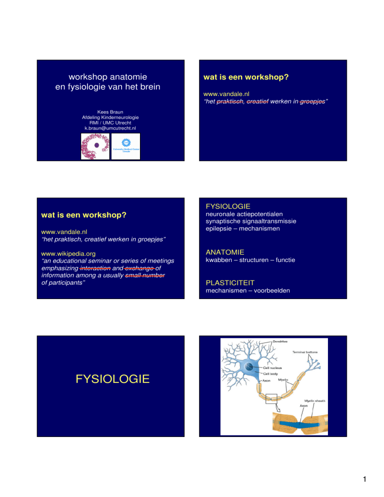
FYSIOLOGIE
wat is een workshop?
www.vandale.nl
“het praktisch, creatief werken in groepjes”
www.wikipedia.org
“an educational seminar or series of meetings
emphasizing interaction and exchange of
information among a usually small number
of participants”
neuronale actiepotentialen
synaptische signaaltransmissie
epilepsie – mechanismen
ANATOMIE
kwabben – structuren – functie
PLASTICITEIT
mechanismen – voorbeelden
FYSIOLOGIE
1
stimulus
response
2
stimulus
stimulus
response
response
stimulus
stimulus
response
response
3
epilepsie:
aanvalsgewijze aandoening
stimulus
response
LTP [long-term potentiation]
epilepsie:
epilepsie:
aanvalsgewijze aandoening
aanvalsgewijze aandoening
>1 aanvallen = (stereotype) verandering
in functie van CZS
>1 aanvallen = (stereotype) verandering
in functie van CZS
t.g.v. onwillekeurige synchrone electrische
ontlading van meerdere neuronen
4
epilepsie:
epilepsie:
aanvalsgewijze aandoening
verlaagde prikkeldrempel neuronen
(hyperexcitabiliteit)
>1 aanvallen = (stereotype) verandering
in functie van CZS
t.g.v. onwillekeurige synchrone electrische
ontlading van meerdere neuronen
structurele of metabole stoornis brein;
diffuus, multifocaal, of focaal
epilepsie:
epilepsie:
verlaagde prikkeldrempel neuronen
(hyperexcitabiliteit)
verlaagde prikkeldrempel neuronen
(hyperexcitabiliteit)
verstoorde balans tussen
excitatoire – inhibatoire neurotransmitters
(glutamaat)
(GABA)
verstoorde balans tussen
excitatoire – inhibatoire neurotransmitters
(glutamaat)
(GABA)
of stoornis in ion-kanalen
epilepsie:
verlaagde prikkeldrempel neuronen
(hyperexcitabiliteit)
verstoorde balans tussen
excitatoire – inhibatoire neurotransmitters
stoornis ionkanalen
overmatige stimulatie neuronen:
- excitotoxiciteit, neuronale schade / dood
5
DWI
DWI
gevolgen van onbehandelde status
epilepsie:
verlaagde prikkeldrempel neuronen
(hyperexcitabiliteit)
verstoorde balans tussen
excitatoire – inhibatoire neurotransmitters
stoornis ionkanalen
overmatige stimulatie neuronen:
- excitotoxiciteit, neuronale schade / dood
- metabole uitputting
epilepsie:
metabole uitputting van neuronen
na epileptische aanval:
postictale
- bewustzijnsdaling
- focale neurologische uitval
- hypofunctie EEG
zenuwgeleiding
synaptische transmissie
neurotransmitters
glutamaat
GABA
ionkanalen
Na+
K+
Ca++
6
zenuwgeleiding
synaptische transmissie
epilepsie
zenuwgeleiding
synaptische transmissie
neurotransmitters
neurotransmitters
glutamaat
GABA
glutamaat
GABA
ionkanalen
ionkanalen
epilepsie
AED
CBZ TPX
VGB VPA
benzo’s TPX
Na+
SCN1A
Na+
SCN1A
K+
Ca++
KCNQ2
CACNA1A
K+
Ca++
KCNQ2
CACNA1A
VPA CBZ
PHT LTG TPX
VPA
ETH
centraal zenuwstelsel
ANATOMIE
functie CZS
opbouw centraal zenuwstelsel
ruggenmerg: baansystemen
• piramidebaan: kracht
• achterstrengen: gnostische sensibiliteit
• tractus spinothalamicus: vitale sensibiliteit
• reflexbogen
• voorhoorncellen
• sympatische banen
opbouw centraal zenuwstelsel
hersenstam (medulla oblongata, pons,
mesencephalon)
•
•
•
•
•
baansystemen
hersenzenuwen, kernen
formatio reticularis
ademhalings- bloeddruk-regulatie centra
blikcentra
“basaal zenuwstelsel”
7
opbouw centraal zenuwstelsel
thalamus
basale kernen (nucleus caudatus, putamen,
globus pallidus)
onderdeel extrapiramidale systeem
extrapiramidaal syndroom:
• stoornis in spiertonus en houding
• onwillekeurige bewegingen
• stoornis in het motorische tempo, balans en
automatiek
opbouw centraal zenuwstelsel
opbouw centraal zenuwstelsel
cerebellum (kleine hersenen)
cortex (hersenschors)
het uitgebalanceerd en gecoördineerd
doen verlopen van bewegingen
ongekruisd
•
•
•
•
stoornis: ataxie (het ontbreken van
geordend bewegen zonder spierzwakte)
bovenaanzicht
EPILEPSIE
frontaalkwab
parietaalkwab
temporaalkwab
occipitaalkwab
in 95% van gevallen: links taaldominant
zijaanzicht
8
binnenaanzicht
onderaanzicht
doorsnede
transversaal
axiaal
coronaal
sagittaal
1
transversaal
coronaal
9
sagittaal
frenologie
functie
10
cortex functie
cortex functie
•
•
•
•
bewustzijn
willekeurige motoriek
sensibiliteit
cognitie:
– taal
– geheugen
– oriëntatie
– planning
– denken
frontaalkwab
gyrus precentralis: primaire motorische cortex
uitvoerende functies
integriteit persoonlijkheid
planning
initiatief
motivatie, inhibitie
area van Broca: taalproductie
11
pariëtaalkwab
gyrus postcentralis: primaire sensorische cortex
visueel ruimtelijke orientatie
rechts: lichaamsschema links
bij stoornis: agnosie, apraxie, neglect
occipitaalkwab
primair visuele cortex
secundair en tertiaire visuele cortex:
visuele herkenning, analyse van plaats,
beweging, kleur, vorm
12
temporaalkwab
area van Wernicke: taalbegrip
primaire en secundaire auditorische
cortex: analyse en directe waarneming geluid
hippocampus: geheugen, leren
nauwe relatie met limbische systeem
emotionele gewaarwordingen
dominante hemisfeer
•
•
•
•
•
taal
lezen
schrijven
rekenen
herkenning
afasie
dyslexie
dysgrafie
dyscalculie
agnosie
niet dominante hemisfeer
•
•
•
•
oriëntatie
aankleden
natekenen
schema lichaam
en ruimte
geografische agnosie
kledingsapraxie
constructieve apraxie
neglect
PLASTICITEIT
13
“is your brain really necessary?”
cerebrale plasticiteit
πλαιστικοσ = vormgeven
vermogen tot verandering en aanpassing
- leren, geheugen, vergeten
- reorganisatie en herstel na schade
handgebied
reorganisatie
15 violisten
35 niet-muzikanten
Schwenkreis et al. 2007
Yu et al. 2006
14
plasticiteit is leeftijds-afhankelijk
kinderen
aanpassing
van de “bedrading”
Büchel et al. 1998
nieuwe verbindingen,
vorming neuronale
netwerken / circuits
>
volwassenen
aanpassing
van de synaps
verandering van
functie en routes in
bestaande netwerken
M1 cortex
stimulus
synaps
spinale
motoneuronen
reactie
M1 cortex
M1 cortex
X
spinale
motoneuronen
X
intracorticale
horizontale
connecties
spinale
motoneuronen
15
M1 cortex
intracorticale
horizontale
connecties
+
-
+
LTP
spinale
motoneuronen
neurogenese
apoptosis
conceptie
aantal
neuronen
geboorte
2jr
dendritische / axonale sprouting
dood
pruning
synaptogenesis
conceptie
geboorte
2jr
dood
aantal
neuronen
conceptie
2jr
aantal
neuronen
synaptische organisatie
conceptie
geboorte
geboorte
2jr
synaptische organisatie
dood
conceptie
geboorte
2jr
dood
verandering
synaps,
corticale
reorganisatie
dood
16
normale ontwikkeling
sensorische input
leren / trainen
omgevingseisen
FYSIOLOGIE
neuronale actiepotentialen
synaptische signaaltransmissie
epilepsie – mechanismen
schade
hersenen
PLASTICITEIT
kritieke periode
aantal
neuronen
synaptische organisatie
verandering
synaps,
corticale
reorganisatie
ANATOMIE
kwabben – structuren – functie
PLASTICITEIT
conceptie
geboorte
2jr
dood
mechanismen – voorbeelden
met dank aan:
Floor Jansen
Onno van Nieuwenhuizen
Frederique van Berkestijn
Ben Vledder
Gerrit-Jan de Haan
17
alter the electrical conditions within and outside the cell membrane.
A nerve cell at rest holds a slight negative charge (about –70 millivolts, or thousandths of a volt, mV) with respect to the
exterior; the cell membrane is said to be polarized. The negative charge, the resting potential of the membrane, arises from
a very slight excess of negatively charged molecules inside the cell.
A membrane at rest is more or less impermeable to positively charged sodium ions (Na+), but when stimulated it is
transiently open to their passage. The Na+ ions thus flow in, attracted by the negative charge inside, and the membrane
temporarily reverses its polarity, with a higher positive charge inside than out. This stage lasts less than a millisecond, and
then the sodium channels close again. Potassium channels (K+) open, and K+ ions move out through the membrane,
reversing the flow of positively charged ions. (Both these channels are known as voltage-gated, meaning that they open or
close in response to changes in electrical charge occurring across the membrane.)
Over the next 3 milliseconds, the membrane becomes slightly hyperpolarized, with a charge of about –80 mV, and then
returns to its resting potential. During this time the sodium channels remain closed; the membrane is in a refractory phase.
An action potential—the very brief pulse of positive membrane voltage—is transmitted forward along the axon; it is
prevented from propagating backward as long as the sodium channels remain closed. After the membrane has returned to
its resting potential, however, a new impulse may arrive to evoke an action potential, and the cycle can begin again.
Gated channels, and the concomitant movement of ions in and out of the cell membrane, are widespread throughout the
nervous system, with sodium, potassium, and chlorine being the most common ions involved. Calcium channels are also
important, particularly at the presynaptic boutons of axons. When the membrane is at its resting potential, positively charged
calcium ions (Ca2+) outside the cell far outnumber those inside. With the advent of an action potential, however, calcium
ions rush into the cell. The influx of calcium ions leads to the release of neurotransmitter into the synaptic cleft; this passes
the signal to a neighboring nerve cell.
Having taken a close look at the electrical side of the picture, we are in a better position to see where the chemistry comes
in. Molecules of neurotransmitter are released into a synaptic cleft and bind to specific receptor sites on the postsynaptic
side (the dendrite or dendritic spine), thereby altering the ion channels in the postsynaptic membrane. Some
neurotransmitters cause sodium channels to open, allowing the influx of Na+ ions and thus a lessening of negative charge
inside the cell membrane. If a considerable number of these potentials are received within a short interval, they can
depolarize the membrane enough to trigger an action potential; the result is the transmission of a nerve impulse. The
substances that can cause this to occur are the excitatory neurotransmitters. By contrast, other chemical compounds cause
potassium channels to open, increasing the outflow of K+ ions from the cell and making excitation less likely; the
neurotransmitters that bring about this state are considered inhibitory.
A given neuron has a great quantity of sites available on its dendrites and cell body and receives signals from many
synapses simultaneously, both excitatory and inhibitory. These signals often amount to a rough balance; it is only when the
net potential of the membrane in one region shifts significantly up or down from the resting level that a particular
neurotransmitter can be said to be exerting an effect. Interestingly, in the membrane's overall balance sheet, the importance
of a particular synapse varies with its proximity to where the axon leaves the nerve cell body, so that numerous excitatory
potentials out at the ends of the dendrites may be overruled by several inhibitory potentials closer to the soma. Other kinds
18


