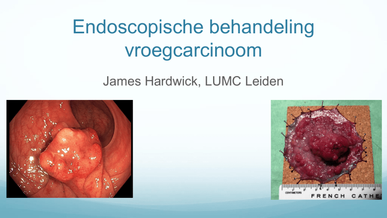
Endoscopische behandeling
vroegcarcinoom
James Hardwick, LUMC Leiden
Early cancer
What is it? What is it not? How often does it occur?
How do you recognise it?
What are the treatment options?
Which endoscopic technique?
When is endoscopic removal sufficient?
What is early cancer?
Early cancer = malignant polyp = ‘cancer’ in a ‘polyp’
Cellular features of cancer
+
Invasion through muscularis mucosae
Biopsies often misleading
What is it NOT?
NOT ‘Carcinoom in situ’ of ‘intramucosale carcinoom’ of ‘
Intra-epitheliaal carcinoom (0% Meta)
Cellular features of cancer but no submucosal invasion
Muscularis mucosae can be difficult to identify
Pseudo-invasion – sigmoid polyps, previous resection
NB pathology tricky - “Expert Board” in UK
How often does it occur?
Screening colonoscopy – 1% incidence
FOBT - ?5% of colonscopies
5% of all polyps
More in larger polyps
More in rectum
Rising incidence
How do you recognise it? - macro
Central depression
Ulceration
Irregular contours
Friability
Smooth surface
Chicken skin
Non-lifting sign
How do you recognise it? - micro
Kudo V pit pattern
How do you recognise it? - micro
Minimise ‘Oeps!’ cancers
Not suspected by endoscopist
Accuracy of recognition (90 vs 50%)
No opportunity to optimise endoscopic resection
Inconclusive/inaccurate histology
Unnecessary surgery
No opportunity to optimally prepare polyp for histology
Mark stalk
Pin sessile/flat polyps to cork
Treatment options?
Colon
Endoscopic resection
Surgical oncological segmental resection
Rectum
Endoscopic
Surgical (TME, APR or LAR)
TEM (SILS port)
Choice for endoscopic resection?
Can and should the lesion be removed endoscopically?
Surgical resection of benign polyp = unnecessary surgery
Endoscopic analysis 50% specific
Endoscopic resection followed by salvage surgery– not detrimental
Poor removal - inconclusive/inaccurate histology, unnecessary surgery
Can I remove it endoscopically?
Time
Scope
Accessories
Experience
Support
Lesions en bloc in one session or refer
Resection or referral?
Referral– excellent photos, accurate estimation of size,
SPOT at a distance, no biopsy, no submucosal
injection
Endoscopic resection– time, positioning of patient,
scoop (retroversion), cap, injection fluid, SPOT
Which endoscopic technique?
Pedunculated - Snare, margin of normal mucosa
Sessile < 2cm - EMR, colloid injection
Sessile < 2cm non-lifting – FTRD, ESD
Sessile >2cm - ESD
EMR
Endoscopic Mucosal Resection
All regions of colon - linea dentata, I-C valve, appendix,
terminal ileum
Quick, safe, easy
No control over depth
Little control over lateral margins
ESD
Endoscopic Submucosal Dissection
Control over lateral and deep margins
Minimises residual/recurrent disease
Optimal specimen for accurate histology
Long procedure time
Higher chance of perforation
Technically very demanding
ESD
Creation of
mucosal flap
ESD
ESD
ESD
6 cm non-granular lesion rectum
Injection of Volufen through polyp
ESD
Incision – not circumferential
Dissection – lesion held taught by sides
ESD
Distal edge of lesion through ‘tunnel’
Resection of edges
ESD
ESD
View in retroversion
Specimen – Small focus of invasive cancer <1mm submucosa
FTRD
Full Thickness Resection Device
Up to 2cm
Technically easy
Damage to tissue outside colon
FTRD
FTRD
FTRD
FTRD
FTRD
FTRD
FTRD
When is endoscopic resection sufficient?
Guidelines:
>1mm resection margin
No lymphangio invasion
No poor differentiation
No Haggit 4
Endoscopic resection sufficient
When is endoscopic resection sufficient?
Guidelines:
Radically removed
No lyphangio invasion
No poor differentiation
Low grade tumour budding
<1000 µM submucosal invasion
Endoscopic resection sufficient
Future
Estimate chance
(%) of residual
cancer
Weigh cancer risk vs operative risk
DICA
Operative risk
Co-morbidity – Charlson score
Age
25
25
20
20
15
15
10
10
5
5
0
0
0
1
2
>=3
UK 1998-2006 30 day mortality
<=50
51-60
61-70
71-80
>=80
Operative risk
American College of Surgeons
National Surgical Quality
Improvement Program
Polypectomy or surgery?
Surgery doesn’t cure all LN+
Morbidity especially rectal surgery
Pathologist and endoscopist must liase
Case history
Pathology – reporting
I: Type biopt :
Polypectomie
Lokalisatie
:
Rectum
Diameter :
3,1
cm
Primaire afwijking :
met invasieve
maligniteit
Snijvlak :
niet vrij
Type
tumor :
Adenocarcinoom
(Lymf)angioinvasie : Niet aanwezig
Differentiatie :
Goed/matig
Invasie diepte :
niet te
beoordelen
Vorm van de laesie:
poliepeus
Pathology – reporting
Revised report
I: Polypectomie Rectum: matig gedifferentieerd
adenocarcinoom met invasieve groei in de steel. Het
adenocarcinoom toont invasieve groei in het bovenste deel
van de poliep en in de steel, derhalve te beschouwen als
kikuchi level 1 tot 2.
Het adenocarcinoom bevindt zich niet in het resectievlak
van de poliep. De tubulair adenoom component met
laaggradige dysplasie reikt wel tot in de basis van de steel,
en is derhalve niet vrij.
Pathology – reporting
Revised revised report
Aanvullend desmine kleuring toont geen ingroei door de muscularis mucosae,
derhalve betreft het een intramucosaal adenocarcinoom. Er is geen invasie in
de submucosa.
I: Poliepectomie rectum, klinisch betreft het een sessiele laesie: intramucosaal
carcinoom, zonder invasie in de submucosa. Het snijvlak van de poliep is vrij
van intramucosaal carcinoom, doch adenoom component met laaggradige
dysplasie reikt tot in het snijvlak. Diameter van de gehele poliep 3,1 cm, geen
lymfangio-invasieve groei. De vorm van de laesie: sessiel. Geen expressie
verlies van de mismatch repair eiwitten. Bovenstaande conclusies vervallen.
Conclusions
Careful endoscopic inspection
Aggressive en bloc resection
Current surgery guidelines unhelpful in the
elderly
No “one size fits all”
Excellent pathology essential – insist on it!
