A diagnostic screening algorithm to asses the public health
risk of STEC.
NVMM Voorjaars vergadering
Poster 029
2013 Papendal
[email protected]
R.F. de Boer1, A. Ott2, H.R. Scheper2, G.J. Wisselink1, J.W. Rossen3, M.E. Heck4, and A.M.D. Kooistra-Smid1,2,3
1 Laboratory for Infectious Diseases, Department of Research & Development, Groningen, the Netherlands.
2 Laboratory for Infectious Diseases, Department of Medical Microbiology, Groningen, the Netherlands.
3 Department of Medical Microbiology, University of Groningen, University Medical Center Groningen, Groningen, the Netherlands
4 National Institute for Public Health and the Environment, Center of Infectious Disease Control, Bilthoven, the Netherlands
Introduction
Results continued
Shiga toxin-producing Escherichia coli (STEC) poses a serious
public health concern because of its ability to cause outbreaks,
hemorrhagic colitis (HC), and hemolytic-uremic syndrome (HUS). In
particular, STEC strains belonging to serotypes O26, O103, O111,
O121, O145 and O157 are known causative agents of outbreaks
and serious illness. This study describes a rapid screening
algorithm to discriminate STEC infections associated with public
health risk from less virulent ones.
• In total, 31/73 samples were confirmed by culture (42%).
• The diagnostic screening algorithm (presumptively) classified
stx1/stx2-pos samples in the following seropathotype (SPT) groups:
SPT A (n=2, 3%)
BRILA PCR
Culture
stx1
stx2
escV
O-type/virulence
Ct
SMAC
CHROM
Serotype
Diagnosis
15
17
16
O157
18
POS
POS
O157:H7
EHEC O157
NEG
29
27
O157/EAEC
31/19
NEG
NEG
SPT B (n=8, 11%)
Materials and Methods
BRILA PCR
• A total of 5022 stool samples from patients with presumed
infectious gastroenteritis were prospectively screened using realtime multiplex PCR (mPCR). Subsequently, PCR positive stool
specimens were cultured directly (SMAC/CHROMagar STEC)
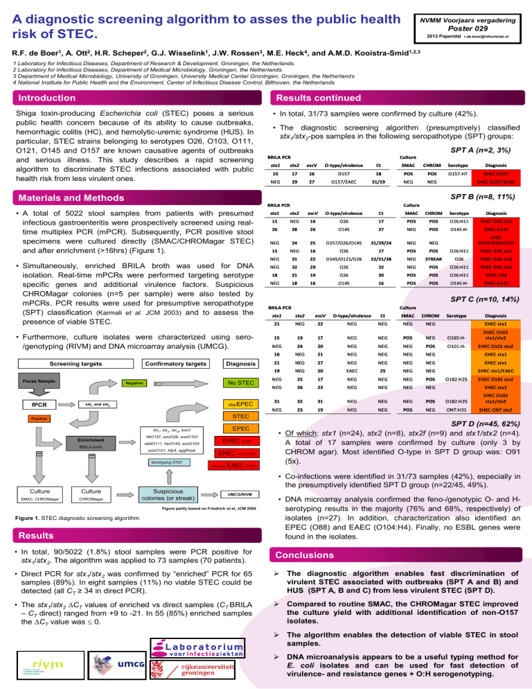
and after enrichment (>16hrs) (Figure 1).
• Simultaneously, enriched BRILA broth was used for DNA
isolation. Real-time mPCRs were performed targeting serotype
specific genes and additional virulence factors. Suspicious
CHROMagar colonies (n=5 per sample) were also tested by
mPCRs. PCR results were used for presumptive seropathotype
(SPT) classification (Karmali et al. JCM 2003) and to assess the
presence of viable STEC.
• Furthermore, culture isolates were characterized using sero/genotyping (RIVM) and DNA microarray analysis (UMCG).
Screening targets
Feces Sample
fPCR
EHEC O157/EAEC
Confirmatory targets
Diagnosis
No STEC
Negative
atypEPEC
stx1 and stx2
Culture
stx1
stx2
escV
O-type/virulence
Ct
SMAC
CHROM
Serotype
Diagnosis
15
NEG
16
O26
17
POS
POS
O26:H11
EHEC O26 stx1
26
28
26
O145
27
NEG
POS
O145:H-
EHEC O145
NEG
24
25
O157/O26/O145
31/29/24
NEG
NEG
EHEC
O145/O26/O157
15
NEG
16
O26
17
POS
POS
O26:H11
EHEC O26 stx1
NEG
31
22
O145/O121/O26
22/31/38
NEG
STREAK
O26
EHEC O26 stx2
NEG
32
29
O26
32
NEG
POS
O26:H11
EHEC O26 stx2
18
21
19
O26
20
POS
POS
O26:H11
EHEC O26
NEG
18
16
O145
16
POS
POS
O145:H-
EHEC O145
SPT C (n=10, 14%)
BRILA PCR
Culture
stx1
stx2
escV
O-type/virulence
Ct
SMAC
CHROM
21
NEG
22
NEG
NEG
NEG
NEG
Serotype
Diagnosis
15
19
17
NEG
NEG
POS
NEG
O165:H-
EHEC O165
stx1/stx2
NEG
24
20
NEG
NEG
NEG
POS
O101:H-
EHEC O101 stx2
16
NEG
21
NEG
NEG
NEG
NEG
21
NEG
27
NEG
NEG
NEG
NEG
EHEC stx1
19
NEG
20
EAEC
25
NEG
NEG
EHEC stx1/EAEC
NEG
25
17
NEG
NEG
NEG
POS
O182:H25
EHEC O182 stx2
NEG
26
23
NEG
NEG
NEG
NEG
EHEC stx2
EHEC stx1
EHEC stx1
31
32
31
NEG
NEG
NEG
POS
O182:H25
EHEC O182
stx1/stx2
NEG
23
19
NEG
NEG
POS
NEG
ONT:H31
EHEC ONT stx2
STEC
Positive
EPEC
stx1, stx2, stx2f, escV
Enrichment
BRILA broth
rfbO157, wzxO26, wzxO103
wbdO111, ihpO145, wzxO104
wzxO121, bfpA, aggR/aat
EHEC O157
EHEC non-O157
Serotyping O157
stx-pos
EAEC O104
SPT D (n=45, 62%)
• Of which: stx1 (n=24), stx2 (n=8), stx2f (n=9) and stx1/stx2 (n=4).
A total of 17 samples were confirmed by culture (only 3 by
CHROM agar). Most identified O-type in SPT D group was: O91
(5x).
• Co-infections were identified in 31/73 samples (42%), especially in
the presumptively identified SPT D group (n=22/45, 49%).
Culture
Culture
SMAC, CHROMagar
CHROMagar
Suspicious
colonies (or streak)
UMCG/RIVM
Figure partly based on Friedrich et al, JCM 2004
Figure 1. STEC diagnostic screening algorithm.
Results
• DNA microarray analysis confirmed the feno-/genotypic O- and Hserotyping results in the majority (76% and 68%, respectively) of
isolates (n=27). In addition, characterization also identified an
EPEC (O88) and EAEC (O104:H4). Finally, no ESBL genes were
found in the isolates.
• In total, 90/5022 (1.8%) stool samples were PCR positive for
stx1/stx2. The algorithm was applied to 73 samples (70 patients).
Conclusions
• Direct PCR for stx1/stx2 was confirmed by “enriched” PCR for 65
samples (89%). In eight samples (11%) no viable STEC could be
detected (all CT ≥ 34 in direct PCR).
The diagnostic algorithm enables fast discrimination of
virulent STEC associated with outbreaks (SPT A and B) and
HUS (SPT A, B and C) from less virulent STEC (SPT D).
• The stx1/stx2 ∆CT values of enriched vs direct samples (CT BRILA
– CT direct) ranged from +9 to -21. In 55 (85%) enriched samples
the ∆CT value was 0.
Compared to routine SMAC, the CHROMagar STEC improved
the culture yield with additional identification of non-O157
isolates.
The algorithm enables the detection of viable STEC in stool
samples.
DNA microanalysis appears to be a useful typing method for
E. coli isolates and can be used for fast detection of
virulence- and resistance genes + O:H serogenotyping.
