Development and evaluation
of DNA amplicon quantification.
Case study: UV–Cabinet with UV Air Recirculator
UVC/T-M-AR and Class II Biological Safety Cabinets
Medical–Biological
Research & Technologies
Authors
Biotechnomica: Marina Tarvida, Julija Isakova,
Vasily Bankovsky
Biosan: Arturs Kigitovics, Vadim Gimelfarb
Introduction
Personal and product safety during clinical and
laboratory studies have stimulated the development of sterile cabinets and special laboratory
safety techniques, to protect the environment,
operator, and product. Monitoring DNA/RNA
amplicon concentration in laboratory air in sterile
cabinets has become topical as PCR and isothermal
amplification technologies have developed along
with wide spread mass analyses.
Development of methods for repeatable
DNA/RNA amplicon detection in air samples is now
a reality. Recent research “Behaviour of aerosol
particles in fibrous structures” (Igor Agranovsky’s
PhD thesis, 2008, Novosibirsk, Russia) describes the
development of samplers and monitoring of DNA/
RNA amplicon concentration in the air from sterile
cabinets, microbial quantitative analyses.
UVC/T-M-AR, UV–Cabinet for PCR operations
110
Applications and Articles
DN
am A/RN
plic A
ons
25 cm
50 cm
ses
Distance to UV source, cm
2 cm
Vir
u
50
ter
ia
2
Bac
25
t
2
4
Yea
s
20
i
Distance,
cm
Fun
g
UV intensity,
mW/cm2/sec
UV intensity needed for decontaminating,
mW/cm2/sec
UV intensity, mW/cm2/sec
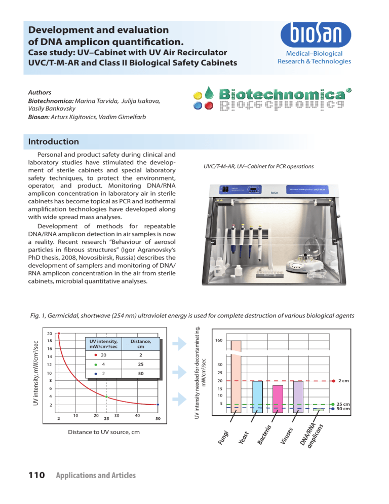
Fig. 1, Germicidal, shortwave (254 nm) ultraviolet energy is used for complete destruction of various biological agents
Aim of the study
Air flow organization through HEPA
filter
The aim of this study is to evaluate the of
efficiency of UV cabinets produced by BioSan
(Latvia) in comparison to Class II BioSafety
cabinets.
HEPA is an acronym for “high efficiency particulate absorbing” or “high efficiency particulate
arrestance” or, as officially defined by the Department of Energy (DOE) “high efficiency particulate
air”.
UV air treatment
More than a century has passed since the
germicidal effect of UV light was recognized by
Niels Ryberg Finsen — a Nobel Prize winner in
physiology or medicine in 1903 [5], and many
researches have been performed on UV induced
destruction of DNA and microorganisms.
The first HEPA filters were developed in the
1940’s by the USA Atomic Energy Commission to
fulfil a an efficient, effective way to filter radioactive particulate contaminants. HEPA filter technology was declassified after World War 2 and
then allowed for commercial and residential use
[6].
Low pressure germicidal UV lamps characteristically emit monochromatic low intensity radiation principally at 253.7 nm, within the germicidal
wavelength range as defined by the DNA absorbance spectrum. The germicidal UV dose LP-UV
lamps is calculated as the product of the volume
averaged incident irradiance (E, mW/cm2) and the
time of exposure (t, seconds) resulting in units of
mJ/cm2 for UV dose [1] (Fig. 1).
This type of air filter can theoretically remove
at least 99.97% of dust, pollen, mold, bacteria
and any airborne particles with a size of 0.3 μm at
85 litres per minute (l/min). In some cases, HEPA
filters can even remove or reduce viral contamination. The diameter specification of 0.3 responds to
the most penetrating particle size (MPPS). Particles that are smaller or larger are trapped with
even higher efficiency [7] (Fig. 2).
Fig. 2, Biological agent sizes and filters effectivity range, nm
HEPA filter
Mechanical Filter
0
100
200
300
600
1 000
2 000
5 000
10 000
DNA/RNA amplicons
Viruses
Bacteria
Yeast
Fungi
Biological agent sizes, nm
Colony forming units (CFU) test
Media
LBA media was prepared using Standard
Methods Agar (Tryptone Glucose Yeast Extract;
Becton, Dickinson and Company) and dissolved in
1 litre of purified water. 7.5 grams of Yeast Extract
(Biolife S.r.l.) and 5 grams of Tryptone (Difco laboratories) were added to enrich the media. The
media was autoclaved at 121°C for 15 minutes.
Media control samples were taken to check for
presence/absence of colony forming units in
media itself and the results were negative (0 CFU
per 3 plates).
Experimental setup:
Impaction aerobiocollector airIDEAL 3P
(bioMérieuxSA, France) was used to take
air samples to test for the presence of colony
forming units (CFU). Each sample was exposed
to 500 litres of air. Aerobiocollector was set in the
middle of the sterile cabinets for test samples and
negative control samples, and in specific places
in the middle of the laboratory room for positive
control. The negative control was taken in Microflow ABS Cabinet Class II. This was repeated three
times, the number of colony forming units was
counted manually on each plate. Reading tables
provided in airIDEAL 3P (bioMérieuxSA, France)
The most probable number (MPN) of microorganisms collected per plate was estimated with
respect to the number of agglomerates of colonies counted on the plate. (MPN was calculated
from the CFU count using FELLER’s law). Subsequently results were converted to CFU per m3.
Applications and Articles
111
Mechanical contamination test
Instrument:
Laser particle counter (produced by Met One,
USA) was used to determine mechanical contamination in the sterile cabinets and laboratory air as
positive control.
Method:
Average amount of particles per litre of air were
measured in sterile cabinet/laboratory air. Meas-
urements were performed 9 times and the average
value presented in the results as number of particles per m3 of air.
Two channels were used to measure amount
of particles of different size: 5 µm and 0.3 µm.
Mechanical filter stops particles larger than 5 µm
while HEPA filter larger then 0.3 µm.
DNA Amplicon test
Instruments:
• Nebulizer, BioSan
• Shaker OS-20, BioSan
• Mini–Centrifuge/Vortex FV-2400, BioSan
• Centrifuge Pico 17, Thermo Electron Corp.
• Centrifuge-Vortex MSC-6000, BioSan
• Real–Time PCR cycler Rotor Gene 3000, Corbett Research
Reagents:
• Lambda DNA, Thermo Fisher Fermentas
• GeneJet Plasmid Miniprep Kit, Thermo
Fisher Fermentas
• Real Time PCR reagents, Central
Research Institute of Epidemiology
Fig. 3, Air and surface samples
and surface sample taking path
Experiment setup:
• Sampling was performed as shown on Fig. 3
• Extraction and analyses were performed as shown on Fig. 4
• Quantitative PCR (Polymerase Chain Reaction):
DNA amplicon quantification in sterile cabinets was
performed by qPCR. Controls and standards were set in each
experiment:
» 4 standards of Lambda DNA of different concentration
prepared in 10 fold dilution: starting concentration
0.6 ng/μl or ≈1,000,000 copies/μl
» 2 NTC (no template control- sterile H2O), experiment was
considered successful only if control was negative.
After samples were taken and extracted as mentioned
above, qPCR reaction master mix was prepared by adding the
following components for each 25 μl of reaction mix to a tube
at room temperature:
x.6
x.5
x.1
x.2
x.7
x.3
x.4
Nebulizer
Samples taken from:
x.1, x.2, x.3 : Air (Syringes)
x.4 : Working surface (Swab)
x.5, x.7 : Side walls (Swabs)
x.6 : Back wall (Swab)
Sample taking path
PCR mix:
2-FL : 7 μl; dNTP’s : 2.5 μl; Forward Primer : 1 μl;
Reverse Primer : 1 μl; DNA probe : 1 μl; Template DNA : 10 μl;
Water, nuclease-free to : 25 μl; Total volume : 25 μl
Table 1, Cycling protocol
Three-step cycling protocol steps
Temperature, °C
Time
Initial denaturation
95
5 min
1
Denaturation
95
5 sec
42
Annealing
60
20 sec
42
Extension
72
15 sec
42
Detection Channel: FAM
112
Applications and Articles
Number of cycles
A
Air /
B
Surface samples
Fig. 4, DNA extraction, samples analyses and result detection
DNA extraction:
A
A
B
From Air Samples :
• Incubation on Shaker OS-20 (BioSan) 180 rpm 15’
• Spin columned (GeneJet Plasmid Miniprep Kit,
Thermo Fisher Fermentas )
B
From Surface Samples:
• Vortex 2-3’’
• Centrifuge at 13,300 rpm for 2’
20 mm
Isolated DNA:
1
2
3
Real time PCR amplification (Fig. 7)
Detection of Ct values and normalization of data (Fig. 8)
Copy number estimation on cabinet volume and surface area
1
2
3
35
30
25
20
15
10
102
103
104
105
106
Results:
Mechanical contamination
Microbial contamination
Results of mechanical air contamination in
cabinets of two types: PCR cabinet (UVC/T-M-AR,
BioSan) and laminar flow cabinets (BioSafety class
II cabinet prototype by BioSan and BSC II cabinet
ABS Cabinet Class II by Microflow) as the positive
control laboratory air samples were taken (Fig. 5).
Microbial contamination in laboratory air and
sterile cabinets. Quantitative results of microbial
air contamination in cabinets of two types: PCR
cabinet (UVC/T-M-AR, BioSan) and laminar flow
cabinets (BioSafety class II cabinet prototype by
BioSan and BSC II cabinet ABS Cabinet Class II by
Microflow) as the positive control laboratory air
samples were taken (Fig. 6).
Fig. 6, Microbial contamination
700
700
600
600
580
600
500
500
CFU/m3
0.3 µm particles n × 103
Fig. 5, Mechanical contamination,
0.3 µm particles
400
300
400
300
200
200
100
100
1
500
0
2
3
Legends for figures 5 and 6:
9
0
1
4
1 Positive control (laboratory air)
2 UV Cabinet (UVC-T-M-AR, Biosan, Latvia)
2
0
3
0
4
3 Laminar flow cabinet
(HEPA BSC II Cabinet prototype, Biosan, Latvia)
4 BSC II Cabinet (ABS Cabinet Class II, Microflow, UK)
Applications and Articles
113
Amplicon contamination-inactivation efficiency:
Results analysis:
Fig. 7, Effect of UV irradiation on Ct/Cq values (raw results)
Real time PCR ensures product quantification using four standards of different
Lambda phage DNA concentration and
comparing Ct/Cq values of samples to
those of concentration standards, based
on standard curve (Fig. 8) (see Corbett
Research Rotor Gene 3000 manual for
more information) Following the amplification Lambda DNA copy number values
were estimated for cabinet volume and
surface area, results presented in (Fig. 9).
Inactivation efficiency was calculated as ratio of DNA amplicons before
and after treatment: direct and indirect
UV treatment for 15 and 30 minutes,
presented in percents in table 2.
Fig. 9, Effect of direct and indirect UV
irradiation on the amplicon concentration
inside PCR cabinet UVC/T-M-AR, Biosan, Latvia
100
100
100
90
1
2
35
30
Ct
80
Relative Copy numbers, %
Fig. 8, Standard curve, influence of
direct and indirect UV irradiation on
lambda phage DNA copy number
25
70
50
40
35
30
20
10
16
8
1
5
0
Air Samples
20
15
Surface Samples
After Lambda phage DNA spraying
10
102
103
104
105
106
UV Air Recirculator for 15 min
(Closed UV light irradiation, 25 W)
Concentration, copy numbers
1
60 60
60
— Samples after Lambda
Phage spraying, no UV
irradiation (positive
control)
— Concentration
standards
2
UV Air Recirculator for 30 min
(Closed UV light irradiation, 25 W)
— Samples after 30 min
UV inactivation
Open UV light (25 W) irradiation for 15 min
Open UV light (25 W) irradiation for 30 min
— Samples
The horizontal axis show: air or surface
samples, along with the relative copy number
presented on vertical axis. Four series represent
inactivation techniques and time of treatment,
open UV light and UV air recirculator treatment
kinetics are presented in the graph.
Table 2. DNA amplicon inactivation efficiency
in PCR cabinet UVC/T-M-AR, Biosan, Latvia
Inactivation method efficiency
Sample
15 min of UV Air Rec.
30 min of UV Air Rec.
15 min of Open UV
+ UV Air Rec.
30 min of Open UV
+ UV Air Rec.
Air Samples
84%
99%
92%
100%
Surface Samples
40%
40%
65%
95%
114
Applications and Articles
Calculation of UV dose for each treatment
Direct UV Irradiation
Fig. 10, UV intensity dependence on distance to UV
tube (measured by radiometer VLX 254, Vilber Lourmat,
France)
Cabinet’s air treatment
BioSan’s cabinet features a single open
UV lamp 25 Watt, germicidal UV irradiation (253.7 nm) measurements have been
performed and UV intensity were recorded
at the level from 20 mW/sec/cm2 to
2 mW/sec/cm2 at distance to UV source from
2 cm to 50 cm respectively. [2] In PCR cabinet
volume following UV intensity gradient
is formed: from 2 mW/cm2 to 20 mW/cm2
(Fig. 10).
UV intensity, mW/cm2/sec
20
UV dosage during treatment = UV intensity
at specific distance (mW/cm2/sec) ×
time of irradiation (sec)
UV dosage during 15 min: gradient from
1,800-18,000 mW/cm2
18
16
14
12
10
8
6
4
2
10
2
20
25
30
40
50
Distance to UV source, cm
UV dosage during 30 min: gradient from
3,600-36,000 mW/cm2
UV intensity,
mW/cm2/sec
Cabinet’s Surface treatment:
Distance to UV source ranges between surfaces
and consequently the UV intensity (table 3):
Distance,
cm
20
2
4
25
2
50
Table 3. Average dosage for different surfaces
Surface
Dosage after 15 min
Dosage after 30 min
Working surface (40-60 cm)
2
1,800-2,700 mW/cm
3,600-5,400 mW/cm2
Side walls (10-60 cm)
1,800-5,400 mW/cm2
3,600-9,000 mW/cm2
Front window (10-60 cm)
1,800-5,400 mW/cm2
3,600-9,000 mW/cm2
UV air recirculation:
Cabinet’s Air treatment
BioSan PCR cabinets feature UV air recirculator. Recirculator consists of a fan, dust filters
and closed UV‑lamp (25 W) installed in a special
aluminium casing, which is located in the upper
hood. Fan's air flow speed is 14 m3/hour, which
processes 1.3 cabinet volumes per minute.
Distance from closed UV lamp to recirculator’s walls is 2 cm at which UV intensity level is
20 mW/sec/cm2 (Fig. 10).
UV air recirculators are designed for constant
air decontamination during operations.
Resulting in following UV dosage for cabinet’s
volume:
• During 15 min recirculation: 380 mW/cm2
• During 30 min recirculation: 780 mW/cm2
Cabinet’s Surface treatment:
UV Air recirculator does not provide cabinet
surface irradiation.
For deactivation of microorganisms and
amplicons on the cabinet’s surface additional
open UV treatment is needed for protection
against contamination
Applications and Articles
115
Conclusions
Air sampling methods developed by
BioSan has been proven to be compatible
with real time PCR detection of product. This
method enables monitoring of laboratory air
and sterile cabinet for presence of target DNA
amplicons.
Based on classification of BioSafety cabinets from European standard EN 12469 [3]
and experiment results: BioSan PCR Cabinets
and Class I, II, III BioSafety Cabinets were
compared on product protection ability in
table 4.
The research was designed to evaluate
BioSan PCR cabinets’ efficiency in comparison
to Class II BioSafety cabinets. Based on the
experiment results PCR cabinets prevent
microbial contamination with inactivation efficiency up to 96%, but in comparison to Class II
BioSafety cabinets do not provide protection
against mechanical contamination.
UV air treatment in BioSan PCR cabinets for
30 min provides DNA amplicon deactivation
efficiency:
• Combined UV treatment (Open UV and
UV air recirculation) provides 100% efficiency
• UV air recirculation provides 99% efficiency
• Open UV irradiation provides 100%
efficiency
Further studies will be focused on:
• Development of high speed monitoring
technology of RNA amplicon concentration
in the laboratory air and in sterile cabinets.
• Investigation of Class II BioSafety cabinets
efficiency against DNA amplicon contamination. Based on preliminary experiment results: DNA amplicon particles which are not
stopped by HEPA filters (Fig. 2) can result in
constant contamination of cabinets volume.
Table 4. Classification of sterile cabinets, based on protection against contamination
Protection against contamination forming units
BioSafety cabinets
Microorganisms
Viruses
DNA/RNA Amplicons
Class I
+
–
–
Class II (A1, A2, B1, B2)
+
–
–
Class III
+
–
–
+/–
+
+
BioSan PCR Cabinets
Table 5. Relation of risk groups to biosafety levels, practices and equipment (source: Laboratory biosafety manual, Third edition)
Risk Biosafety
Group Level
Laboratory
Type
Laboratory
Practices
Safety
Equipment
1
Basic — Biosafety
Level 1
Basic teaching, research
GMT
None; open bench work
2
Basic — Biosafety
Level 2
Primary health services;
diagnostic services, research
GMT plus protective clothing,
biohazard sign
Open bench plus BSC for potential
aerosols
3
Containment —
Biosafety Level 3
Special diagnostic services,
research
As Level 2 plus special clothing,
BSC and/or other primary devices for all
controlled access, directional airflow activities
4
Maximum Containment Dangerous pathogen units
— Biosafety Level 4
As Level 3 plus airlock entry, shower Class III BSC or positive pressure suits in
exit, special waste disposal
conjunction with Class II BSCs, doubleended autoclave (through the wall),
filtered air
BSC, biological safety cabinet; GMT, good microbiological techniques
116
Applications and Articles
Table 6. Summary of biosafety level requirements (source: Laboratory biosafety manual, Third edition)
Biosafety Level
1
2
3
4
Isolation a of laboratory
No
No
Yes
Yes
Room sealable for decontamination
No
No
Yes
Yes
— inward airflow
No
Desirable
Yes
Yes
— controlled ventilating system
No
Desirable
Yes
— HEPA-filtered air exhaust
No
No
Yes/No
Double-door entry
No
No
Yes
Yes
Airlock
No
No
No
Yes
Airlock with shower
No
No
No
Yes
Anteroom
No
No
Yes
—
Anteroom with shower
No
No
Yes/No c
No
Effluent treatment
No
No
Yes/No
Yes
— on site
No
Desirable
Yes
Yes
— in laboratory room
No
No
Desirable
Yes
— double-ended
No
No
Desirable
Yes
Biological safety cabinets
No
Desirable
Yes
Yes
No
No
Desirable
Yes
Ventilation:
Yes
b
c
Yes
Autoclave:
Personnel safety monitoring capability
d
Environmental and functional isolation from general traffic.
Dependent on location of exhaust (see Chapter 4 of Laboratory Biosafety Manual).
c Dependent on agent(s) used in the laboratory.
d For example, window, closed-circuit television, two-way communication.
a b Acknowledgement
We acknowledge BioSan for financial support
and technical assistance, Anete Dudele for work
done in the beginning of the research on microbial contamination in PCR cabinets.
We acknowledge Central Researcha Institute of
Epidemiology (Moscow, Russia) and M. Markelov,
G. Pokrovsky, and V. Dedkov in particular, for
development and provision reagents for lambda
DNA quantitative analysis using Real–Time PCR
method.
We acknowledge Paul Pergande for donating
his time and expertise by reviewing this article.
References
1. K Linden, A Mofidi. 2004. Disinfection Efficiency and
Dose Measurement of Polychromatic UV Light (1-6)
2. BioSan UV-air flow Cleaner-Recirculators test
report (http://www.biosan.lv/eng/uploads/images/
uvrm%20uvrmi%20article%20eng.pdf )
3. European Committee for Standardization (2000)
European standard EN 12469: BiotechnologyPerformance criteria for microbiological safety
cabinets.
4. Web source: http://nobelprize.org
5. Web source: http://www.aircleaners.com/
hepahistory.phtml
6. Web source: http://www.filt-air.com/Resources/
Articles/hepa/hepa_filters.aspx#Characteristics
7. Web source: http://www.who.int/csr/resources/
publications/biosafety/Biosafety7.pdf
8. Laboratory biosafety manual, Third edition
Applications and Articles
117


